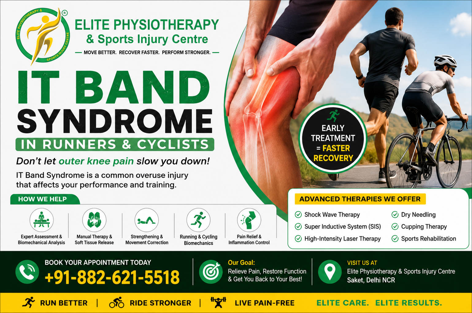
Among the most well-liked endurance sports are cycling and running. However, the lower limbs may be severely stressed by repetitive training. Iliotibial Band Syndrome (ITBS) is a common overuse injury that affects both cyclists and runners. This disease is a prevalent cause of lateral knee pain and can drastically decrease athletic performance.
IT Band Syndrome Physiotherapy plays a significant role in relieving discomfort, correcting biomechanical flaws, restoring movement, and enabling athletes return to sport safely. Early intervention is critical since untreated symptoms can become chronic and compromise training consistency.
We frequently evaluate and treat runners, cyclists, triathletes, and recreational athletes with IT Band Syndrome at Elite Physiotherapy and Sports Injury Centre in Delhi NCR. To achieve the best possible recovery, our all-inclusive rehabilitation programs incorporate cutting-edge technologies, strength training, movement correction, and modern physiotherapy techniques.
Whether you’re looking for physiotherapy Saket, physiotherapy Delhi, or physiotherapy near me, knowing what IT Band Syndrome is can help you get well.
What Is IT Band Syndrome?
A thick, fibrous band of connective tissue that runs from the outside of the pelvis to the outside of the tibia is known as the iliotibial band (IT Band). The tensor fascia lata and gluteus maximus muscles join to it.
The IT band assists:
Maintain knee stability during running and walking.
Help with hip movement
Regulate the position of your lower limbs
Boost the transmission of force when participating in sports
The tissues surrounding the lateral femoral epicondyle are impacted by excessive friction, compression, or repetitive stress, which results in Iliotibial Band Syndrome (ITBS). The outside of the knee becomes painful as a result.
The condition commonly develops in:
Long-distance runners
Cyclists
Triathletes
Hikers
Military personnel
Athletes participating in repetitive knee flexion activities
Understanding the Mechanism of Injury
Previously, ITBS was considered a friction syndrome caused by the IT band repeatedly rubbing over the lateral femoral epicondyle.
According to available data, compression of highly innervated tissues beneath the IT band may be a significant factor. Running and cycling cause repetitive knee bending, which puts more strain on these structures.
Consequently:
There is localized inflammation.
Increased tissue irritation
Pain happens while engaging in an activity.
Performance gradually deteriorates
Epidemiology of IT Band Syndrome
One of the most prevalent overuse injuries among endurance athletes is IT Band Syndrome.
Studies reveal that:
ITBS accounts for a significant percentage of running injuries.
It is among the leading causes of lateral knee pain.
Distance runners show higher incidence rates.
Cyclists frequently develop symptoms due to repetitive pedaling mechanics.
Athletes of all ages and skill levels may be impacted by the condition .
Causes of IT Band Syndrome
Training Errors (Sudden mileage increase, Rapid increase in cycling volume, Excessive hill running, Inadequate recovery, High training frequency)
Many athletes continue training despite symptoms. Unfortunately, this often worsens tissue irritation.
Early IT Band Syndrome Physiotherapy can:
Reduce pain quickly
Correct biomechanical faults
Prevent chronic symptoms
Improve athletic performance
Reduce recurrence risk
Enable a safe return to sport
At Elite Physiotherapy and Sports Injury Centre, Delhi NCR, we use evidence-based assessment methods and advanced rehabilitation protocols to identify the root cause of IT Band Syndrome rather than simply treating symptoms.
IT Band Syndrome Physiotherapy & Rehabilitation Guide
Successful treatment requires more than pain relief. The primary goal is to identify and correct the underlying cause of the condition. At Elite Physiotherapy and Sports Injury Centre, Delhi NCR, we focus on restoring normal biomechanics, improving strength, optimizing movement patterns, and enabling a safe return to running and cycling.
Goals of IT Band Syndrome Physiotherapy
Reduce pain and inflammation
Improve tissue healing
Restore normal movement
Improve flexibility
Correct biomechanical faults
Enhance hip and core strength
Improve running mechanics
Improve cycling biomechanics
Prevent recurrence
Return athletes safely to sport
Pain Reduction and Tissue Recovery
Initial Activity Modification
Temporary reduction in running volume
Shorter cycling sessions
Avoidance of downhill running
Avoidance of excessive hill training
Reduced training intensity
Pain Management Strategies
Ice application
Relative rest
Manual therapy
Therapeutic modalities
Soft tissue techniques
Manual Therapy Techniques
Soft Tissue Mobilization
Myofascial Release
Joint Mobilization
Stretching
Tensor Fascia Lata Stretch
Hip Flexor Stretch
Gluteal Stretch
Hamstring Stretch
Calf Stretch
Strengthening
Hip Abductor Strengthening
Hip Extensor Strengthening
Core Stability Training
Quadriceps Strengthening
Neuromuscular Re-Education
Single-leg balance
Dynamic balance drills
Step-down retraining
Landing mechanics
Agility exercises
Running Retraining
Cycling Biomechanics Correction
Advanced Physiotherapy Modalities for IT Band Syndrome
Shock Wave Therapy
Super Inductive System (SIS) Therapy
High-Intensity Laser Therapy
Dry Needling for IT Band Syndrome
Cupping Therapy
Return-to-Sport Criteria
Full knee range of motion
Minimal or no pain
Normal hip strength
Good single-leg control
Pain-free squatting
Pain-free running or cycling drills
Improved movement mechanics
Prevention of IT Band Syndrome
Training Load Management
Strength Training
Mobility Maintenance
Proper Footwear
Bike Fit Optimization
Why Choose Elite Physiotherapy and Sports Injury Centre for IT Band Syndrome Physiotherapy?
At Elite Physiotherapy and Sports Injury Centre, Delhi NCR, we specialize in the assessment and treatment of sports injuries affecting runners, cyclists, and endurance athletes.
Our approach combines:
Comprehensive biomechanical assessment
Sports-specific physiotherapy
Running gait analysis
Advanced rehabilitation protocols
Shock Wave Therapy
Super Inductive System (SIS)
High-Intensity Laser Therapy
Dry Needling
Cupping Therapy
Return-to-sport testing
Whether you are searching for physiotherapy near me, physiotherapy Delhi, or physiotherapy Saket, our team provides individualized care designed to restore performance and prevent future injuries.
Conclusion
One of the most frequent overuse ailments among cyclists and runners is IT Band Syndrome. But pain management alone is not enough for a full recovery. Strength deficiencies, movement dysfunctions, training mistakes, and biomechanical flaws must all be addressed by athletes.
An organized program of physical therapy for IT Band Syndrome can greatly speed up recovery, lower the chance of recurrence, and improve sports performance. Long-term success is still largely dependent on early diagnosis and evidence-based rehabilitation.
With cutting-edge physiotherapy and sports rehabilitation techniques, we at Elite Physiotherapy and Sports Injury Centre, Delhi NCR, help athletes recover stronger, move more effectively, and perform at their peak.
Patellar Tendinitis Physiotherapy: The Key to a Stronger Comeback
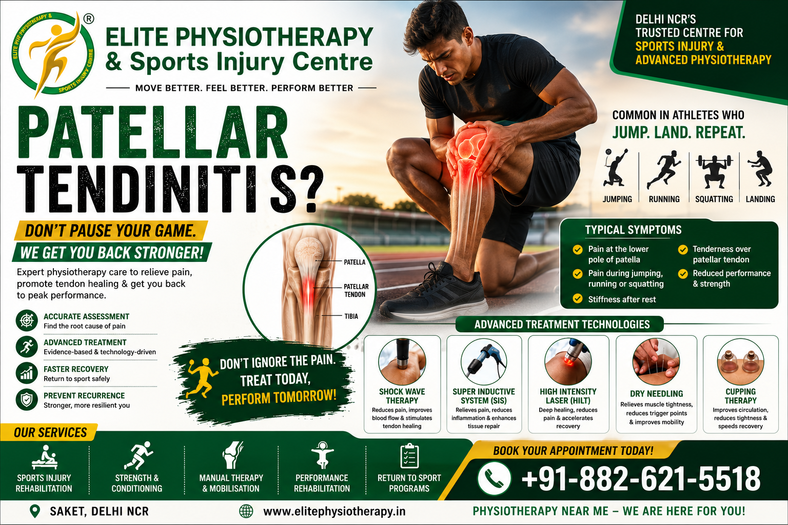
Athletes continuously challenge their bodies to reach their maximum potential. However, the knee joint is severely stressed by frequent jumping, sprinting, landing, and abrupt direction changes. Because of this, a lot of athletes get patellar tendinitis, also referred to as Jumper’s Knee.
We regularly treat athletes with this problem at Elite Physiotherapy and Sports Injury Centre, Delhi NCR. Athletes can safely recuperate and resume their best performance with the aid of our evidence-based rehabilitation programs.
Patellar tendinitis physiotherapy is the best healing method. Early management enhances tendon repair, restores strength, lessens pain, and stops recurrence.
Knowing this problem will help you seek prompt treatment, whether you are a fitness enthusiast, professional athlete, or leisure athlete looking for physiotherapy near me.
What is Patellar Tendinitis?
An overuse injury that affects the patellar tendon is called patellar tendinitis. The patella, or kneecap, and tibia, or shinbone, are joined by this tendon.
The patellar tendon plays a critical role during:
Running
Sprinting
Jumping
Landing
Squatting
Kicking
The tendon sustains microscopic injury as a result of repeated stress. Degeneration occurs when the tendon is unable to recover sufficiently in between training sessions. Pain and decreased athletic performance follow.
While the word “tendinitis” implies inflammation, tendon degradation is more common in chronic instances than active inflammation. As a result, tendon loading and tissue remodeling are the main goals of contemporary rehabilitation.
Understanding Patellar Tendinitis Physiotherapy
Restoring tendon health while addressing the underlying causes of overload is the goal of patellar tendinitis physical therapy.
The main goals of treatment are:
Pain reduction
Tendon healing
Strength restoration
Biomechanical correction
Return-to-sport preparation
Injury prevention
We create customized rehabilitation plans at Elite Physiotherapy and Sports Injury Centre depending on the athlete’s sport, training requirements, mobility patterns, and recuperation objectives.
Who Commonly Develops Patellar Tendinitis?
This condition is frequently seen in:
Volleyball players
Basketball players
Football players
Wrestlers
Weightlifters
Track and field athletes
Badminton players
Tennis players
CrossFit athletes
Athletes involved in explosive jumping and landing activities are particularly vulnerable.
Causes of Patellar Tendinitis
Repetitive Jumping Activities
Sudden Increase in Training Load
Muscle Weakness
Poor Landing Mechanics
Reduced Flexibility
Training Surface Issues
Biomechanical Abnormalities
Clinical Features and Symptoms
Anterior Knee Pain
Pain During Jumping
Pain During Squats
Morning Stiffness
Tenderness
Reduced Performance
Swelling
Patellar Tendinitis Physiotherapy Management
For the majority of athletes, patellar tendinitis physiotherapy is still the best course of action.
A methodical and systematic strategy is necessary for successful rehabilitation.
Our programs at Elite Physiotherapy and Sports Injury Centre are customized based on:
Sport demands
Stage of injury
Pain severity
Performance goals
Pain Reduction and Load Management
The initial goal is symptom control. However, complete rest is rarely recommended. Instead, we modify activities while maintaining fitness.
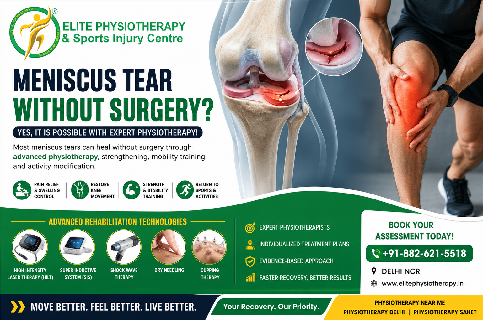
Every facet of daily living can be impacted by knee discomfort. Running, crouching, walking, climbing stairs, and participating in sports may become challenging. Meniscus injuries are among the most frequent causes of knee discomfort. Thankfully, surgery is not always required. Under the direction of skilled physiotherapists, Meniscus Tear Without Surgery is frequently a practical and effective therapy strategy.
Using evidence-based rehabilitation methods, we at Elite Physiotherapy & Sports Injury Centre frequently assist patients and athletes in recovering from meniscus injuries. Our objectives are to lessen discomfort, enhance strength, restore knee function, and enable people to safely resume their intended activities.
Knowing the function of physiotherapy in meniscus recovery will help you make wise choices whether you’re looking for physiotherapy, physiotherapy Delhi, physiotherapy Saket, physiotherapy near me, or specialized sports rehabilitation services.
What Is a Meniscus Tear?
Between the shin bone (tibia) and the thigh bone (femur) is a C-shaped cartilage structure called the meniscus. There are two menisci in each knee:
Medial meniscus (inside)
Lateral meniscus (outside)
Menisci serve as shock absorbers. They increase joint stability and disperse load across the knee joint. Additionally, they aid in preventing excessive stress on the articular cartilage.
When this cartilage is harmed by abrupt twisting, high loading, degeneration, or trauma, a meniscus tear results.
A lot of folks think that surgery is the only option. However, current research indicates that for certain patients, meniscus tears without surgery can be quite beneficial.
Can a Meniscus Tear Heal Without Surgery?
The response is contingent upon multiple factors:
Tear location
Tear size
Patient age
Activity level
Knee stability
Presence of locking symptoms
The blood supply is better in the meniscus’s outer region. As a result, tears in this area frequently heal more quickly.
Structured physiotherapy works incredibly well for many small-to-moderate rips. Without surgery, degenerative meniscus rips often heal.
Whether you are searching for physiotherapy Delhi, physiotherapy Saket, elite rehabilitation services, or physiotherapy near me, our team is committed to helping you move pain-free and return to the activities you enjoy.
Conclusion
Surgery is not always necessary when a meniscus tear occurs. A well-designed physiotherapy program can often prevent meniscus tears without the need for surgery. Knee function and quality of life can be greatly enhanced by appropriate evaluation, gradual strengthening, mobility restoration, and functional therapy.
Seek a professional checkup if you are having knee pain, locking, or trouble playing sports. Better results and quicker recovery are frequently the results of early intervention.
One of the most frequent complaints from runners and other athletic people is knee discomfort. Runner’s Knee Physiotherapy Treatment is one of the most sought-after rehabilitation options among all knee-related issues since it can impact daily activities, sports performance, and training.
We frequently treat athletes, runners, fitness enthusiasts, and active people with runner’s knee at Elite Physiotherapy & Sports Injury Centre. To assist patients in resuming pain-free activities, our evidence-based approach integrates comprehensive evaluation, cutting-edge physiotherapy methods, strength training, movement correction, and contemporary rehabilitation technologies.
Understanding this disease is the first step toward rehabilitation, regardless of whether you are looking for physiotherapy in Saket, Delhi, or your area.
What is Runner’s Knee?
Patellofemoral Pain Syndrome (PFPS) is commonly referred to as “runner’s knee.” It describes discomfort behind or around the patella, or kneecap. When the patella does not move in the femoral groove as it should during movement, discomfort results.
Although the name suggests it only affects runners, the condition is also common among:
Athletes
Cyclists
Football players
Weightlifters
Hikers
Gym-goers
Individuals with poor lower limb mechanics
The pain typically increases during activities that place repeated stress on the patellofemoral joint.
Types and Classification of Runner’s Knee
Runner’s knee can be classified according to contributing factors.
Overuse-Related Runner’s Knee
This develops due to repetitive loading without adequate recovery.
Biomechanical Runner’s Knee
This occurs because of abnormal movement patterns, muscle imbalance, or poor alignment.
Muscular Imbalance Related Runner’s Knee
Weakness of specific muscle groups causes altered patellar tracking.
Structural Runner’s Knee
Structural abnormalities may contribute to patellofemoral joint stress.
Causes of Runner’s Knee
Training Errors
Muscle Weakness
Poor Patellar Tracking
Hip Dysfunction
Foot and Ankle Issues
Reduced Flexibility
Previous Injury
Signs and Symptoms
Pain around the kneecap
Pain behind the kneecap
Pain during running
Pain while climbing stairs
Pain while descending stairs
Pain during squatting
Pain after prolonged sitting
Knee stiffness
Clicking sensations
Reduced athletic performance
Many patients report discomfort during activities involving repeated knee bending.
Physiotherapy Treatment at Elite Physiotherapy
Instead of only treating symptoms, Elite Physiotherapy & Sports Injury Center aims to address the underlying problem.
Pain Management
Activity modification
Manual therapy
Taping techniques
Soft tissue release
Joint mobilization
Patellar Tracking Correction
Patellar mobilization
Taping techniques
Muscle activation training
Strength Training for
Gluteus Medius
Gluteus Maximus
Quadriceps
Hamstrings
Calf Muscles
Core Muscles
Mobility Restoration
Hip mobility exercises
Ankle mobility exercises
Quadriceps stretching
Hamstring stretching
Calf stretching
IT band mobility techniques
Movement Re-Education
Walking mechanics
Running technique
Landing mechanics
Squatting patterns
Single-leg control
Running Retraining
Step rate
Stride length
Foot strike pattern
Hip mechanics
Knee loading patterns
Sport-Specific Rehabilitation
Agility drills
Plyometric training
Strength testing
Functional testing
Return-to-sport preparation
Advanced Modalities Used at Elite Physiotherapy & Sports Injury Centre
Instead than treating symptoms, our team concentrates on determining the underlying cause of discomfort. To provide long-lasting benefits, we integrate professional physiotherapy, cutting-edge technology, sports rehabilitation, strength and conditioning, and movement analysis.
Whether you’re an athlete, recreational runner, or fitness enthusiast, our mission is to improve your mobility, speed up your recovery, and make a stronger comeback.
Our knowledgeable staff is prepared to assist you if you’re searching for physiotherapy in Delhi, Saket, Elite, or around.
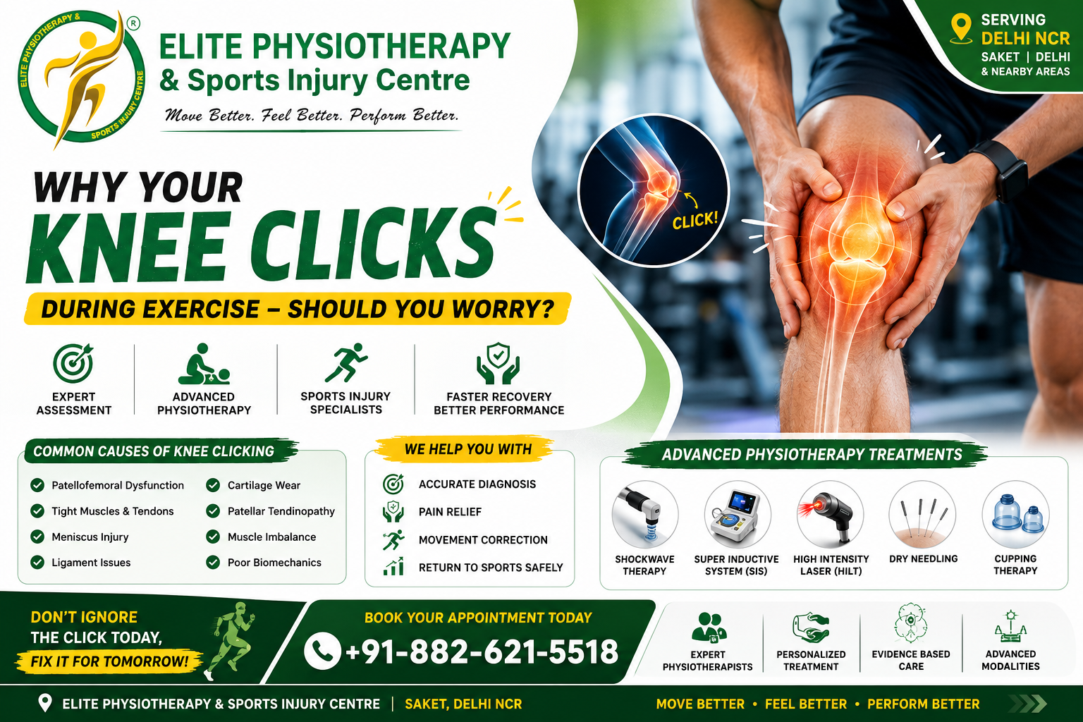
Should You Worry About Knee Clicking During Exercise?
During exercise, a lot of people hear odd noises coming from their knees. Running, stair climbing, squats, and other gym workouts might occasionally cause the knee to pop, fracture, or click. These sounds are often innocuous. However, some people report that the sound is accompanied by pain, swelling, or instability. Consequently, it becomes crucial to comprehend the cause of knee clicking during exercise.
We regularly evaluate athletes, fitness enthusiasts, runners, office workers, and gym patrons with knee issues at Elite Physiotherapy & Sports Injury Centre. Patients in Delhi NCR can receive evidence-based physiotherapy treatment regimens from our knowledgeable staff. Whether you’re looking for physiotherapy Saket, physiotherapy Delhi, or physiotherapy near me, our clinic focuses on treating the underlying problem rather than just the symptoms.
The causes, symptoms, diagnosis, physiotherapy evaluation, and advanced rehabilitation techniques for knee clicking during activity are all covered in this comprehensive book.
What Is Knee Clicking During Exercise?
The term “knee clicking” describes auditory or tactile noises produced by the knee joint during motion.
These noises could consist of:
Clicking
Popping
Cracking
Snapping
Grinding
This ailment is frequently referred to as “crepitus” by medical professionals. Certain clicking sounds are produced spontaneously by soft tissue movement, gas bubbles, or joint movement. On the other hand, chronic or painful clicking could be a sign of ligament damage, cartilage irritation, muscular imbalance, or joint dysfunction.
The knee joint is very intricate. Bones, cartilage, ligaments, tendons, muscles, and synovial structures are all included. As a result, even little movement mistakes can produce strange noises.
Is Knee Clicking Dangerous?
Not all the time.
In most cases, painless knee clicking is not dangerous. When squatting or ascending stairs, a lot of healthy people click without getting hurt. However, you should get evaluated by a physiotherapist if the clicking is connected to:
Pain
Swelling
Knee locking
Instability
Reduced motion
Difficulty exercising
Recent trauma
Recurrent giving way
To determine if the clicking is pathological or normal, Elite Physiotherapy & Sports Injury Center always conducts thorough biomechanical investigation.
Common Causes of Knee Clicking During Exercise
Patellofemoral Joint Dysfunction
When moving, the kneecap could not glide smoothly. Friction between the patella and femur results from this. When squatting or climbing stairs, this frequently makes clicking noises.
This condition is very prevalent among gym-goers and runners.
Contributing factors:
Weak quadriceps
Tight iliotibial band
Poor hip control
Muscle imbalance
Flat feet
Tight Muscles and Tendons
During movement, tendons can occasionally break across bone surfaces. Abnormal tracking may be caused by tight quadriceps, hamstrings, or the iliotibial band.
This is a common reaction of athletes to intense training loads.
Meniscus Injury
Inside the knee, the meniscus serves as a shock absorber. Clicking, catching, or locking sensations could be caused by a torn meniscus.
Common symptoms:
Joint line pain
Swelling
Locking
Pain during twisting
This issue is frequently brought on by sports injuries.
Ligament Injury
Knee mechanics may be affected by partial ligament damage. During exercise, clicking sounds may result from abnormal movement patterns.
Commonly involved ligaments include:
ACL
PCL
MCL
Cartilage Wear and Early Arthritis
Friction within the knee is increased by cartilage degradation. Commonly involved ligaments include:As a result, clicking and grinding noises could appear.
This is more common in:
Older adults
Obese individuals
Previous knee injury patients
Synovial Plica Syndrome
The knee joint contains a fold called the synovial plica. Sensations of snapping or clicking may result from irritation of this structure.
Athletes who frequently bend their knees are particularly vulnerable.
Patellar Tendinopathy
When jumping or landing, overuse of the patellar tendon can cause pain and clicking.
This condition is common in:
Basketball players
Volleyball athletes
Runners
Gym athletes
Gas Bubble Formation
Gas bubbles can occasionally occur in joint fluid. There is a bursting sound when these bubbles explode. Usually, this is harmless and painless.
Types of Knee Clicking
Physiological Clicking
This type is harmless.
Features:
No pain
No swelling
Full movement
Occasional sound only
Pathological Clicking
This requires medical assessment.
Features:
Painful clicking
Swelling
Locking
Instability
Reduced performance
Common risk factors:
Poor exercise technique
Muscle weakness
Sudden training increase
Previous knee injury
Obesity
Sedentary lifestyle
Tight muscles
Improper footwear
Poor biomechanics
Weak hip muscles
Physiotherapy Management for Knee Clicking During Exercise
Physiotherapy is the most effective conservative treatment for most knee clicking conditions.
At Elite Physiotherapy & Sports Injury Centre, treatment focuses on:
Reducing pain
Improving movement quality
Correcting biomechanics
Restoring muscle balance
Preventing recurrence
Returning patients to exercise safely
Pain Management and Activity Modification
Initially, painful activities are modified temporarily.
However, complete rest is rarely recommended. Controlled movement promotes healing and maintains strength.
Elite Physiotherapy & Sports Injury Centre provides advanced evidence-based rehabilitation across Delhi NCR.
Our clinic combines:
Sports physiotherapy expertise
Biomechanical analysis
Manual therapy
Advanced modalities
Athlete rehabilitation
Return-to-play testing
Whether you are searching for physiotherapy near me, physiotherapy Delhi, or physiotherapy Saket, our expert team delivers personalized rehabilitation programs for every patient.
We focus on restoring movement, reducing pain, and helping you return to activity confidently.
Conclusion
Exercise-related knee clicking is widespread. Thankfully, a large number of instances are benign. But clicking that hurts or doesn’t go away should never be disregarded.
The precise cause might be determined with the aid of a thorough physiotherapy evaluation. Early intervention promotes long-term knee health and stops symptoms from getting worse.
Our skilled physiotherapists at Elite Physiotherapy & Sporting Injury Centre provide individualized rehabilitation plans based on your functional objectives, sporting demands, and movement patterns.
Don’t wait for your knee to get worse if it clicks while you’re exercising. Your performance can be safely enhanced and pain-free movement restored with the right physiotherapy treatment.
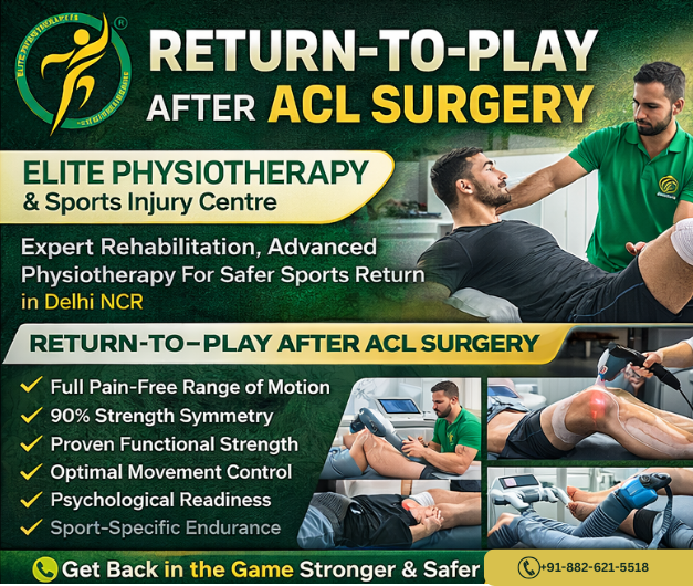
Athletes and active people frequently have anterior cruciate ligament injuries. Surgery by itself, however, does not ensure a safe return to sports. Return-to-Play Requirements: There is a methodical decision-making process following ACL surgery. It guarantees that before the competition, the athlete regains strength, stability, and confidence.
Movement quality, performance readiness, and scientific rehabilitation are our main priorities at Elite Physiotherapy and Sports Injury Centre. Our cutting-edge physiotherapy method in Delhi NCR lowers the chance of reinjury and helps athletes return to their best.
The objective is straightforward. Make a stronger, safer, and more intelligent return.
What is ACL Surgery Rehabilitation?
ACL restoration uses a graft to replace the damaged ligament. Knee function is then gradually restored through rehabilitation.
Return-to-Play Criteria After ACL Surgery refers to meeting specific functional and physical requirements before engaging in sports.
Readiness is not just determined by time. Functional recovery does.
Causes of ACL Injury
Sudden direction change
Improper landing mechanics
Knee valgus collapse
Weak hip and core muscles
Poor neuromuscular control
Previous knee injury
Athletes in football, wrestling, basketball, and badminton face a higher risk.
Types of ACL Reconstruction
Classification depends on graft selection:
Hamstring tendon graft
Patellar tendon graft
Quadriceps tendon graft
Allograft reconstruction
Each type requires a slightly modified physiotherapy progression.
Clinical Features After ACL Surgery
Patients may experience:
Knee swelling
Quadriceps weakness
Reduced range of motion
Instability feeling
Pain during loading
Fear of movement
Therefore, structured Return-to-Play Criteria After ACL Surgery becomes essential.
Diagnostic Evaluation and Examination
A detailed assessment guides rehabilitation progression.
Clinical Examination Includes:
Range of motion assessment
Swelling evaluation
Muscle strength testing
Movement analysis
Functional performance testing
Assessment Techniques for Return-to-Play
At Elite Physiotherapy, assessment remains objective and measurable.
1. Strength Testing
Quadriceps and hamstring strength should reach at least 90% symmetry compared to the opposite limb.
2. Functional Hop Tests
Single hop for distance
Triple hop test
Crossover hop test
Timed hop test
Poor symmetry indicates incomplete recovery.
3. Movement Quality Analysis
Psychological Readiness
Physiotherapy Management
Physiotherapy forms the foundation of Return-to-Play Criteria After ACL Surgery. Rehabilitation progresses through phases.
Phase 1: Protection and Activation
Early physiotherapy focuses on swelling reduction and mobility.
Treatment includes:
Cryotherapy
Patellar mobilization
Gentle range exercises
Quadriceps activation drills
Gait correction training
Early muscle activation prevents long-term weakness.
Phase 2: Strength Restoration
Once pain reduces, progressive strengthening begins.
Key exercises:
Closed chain strengthening
Controlled squats
Step-ups
Hip and core stability training
At our physiotherapy facility in Saket & gurugram, movement quality takes priority over load.
Phase 3: Neuromuscular Control
This phase rebuilds coordination and joint awareness.
Training includes:
Balance training
Proprioceptive drills
Perturbation exercises
Single-leg stability work
These exercises prepare the knee for unpredictable sports movements.
Phase 4: Power and Plyometric Training
Athletes begin explosive training gradually.
Programs include:
Jump mechanics correction
Deceleration drills
Agility ladder work
Sport-specific movement retraining
This stage strongly influences the Return-to-Play Criteria After ACL Surgery success.
Phase 5: Sports Reintegration
Final physiotherapy focuses on performance.
We introduce:
Cutting drills
Sprint progression
Reaction training
Match simulation exercises
Athletes must pass objective testing before clearance.
Role of Advanced Physiotherapy Modalities
Shock Wave Therapy
Improves tissue healing and reduces persistent tendon pain around the knee.
Super Inductive System (SIS)
Enhances neuromuscular activation. It improves quadriceps recruitment safely.
High Intensity Laser Therapy
Reduces inflammation and promotes deep tissue repair.
Dry Needling and Cupping
Relieves muscle tightness and improves circulation. These methods reduce compensatory movement patterns.
These modalities support faster and safer rehabilitation when combined with exercise therapy.
Key Return-to-Play Criteria After ACL Surgery
Athletes must achieve:
Full pain-free range of motion
Minimal swelling
Strength symmetry above 90%
Proper landing mechanics
Successful hop test performance
Psychological readiness
Sport-specific endurance
Meeting all criteria lowers reinjury risk significantly.
Why Choose Elite Physiotherapy and Sports Injury Centre?
We deliver evidence-based physiotherapy near me solutions across Delhi NCR.
Our approach includes:
Personalized rehabilitation programs
Athlete-specific return protocols
Advanced technology modalities
Performance testing systems
Injury prevention education
At Elite, we do not rush returns. We prepare athletes for long careers.
Conclusion
It’s not a race to recover from an ACL. It is a methodical process.
Athletes can return to play with confidence and safety thanks to the Return-to-Play Criteria following ACL surgery. Effective physiotherapy turns recovery into improved performance.
Do you get knee pain when you squat during daily activities or workouts? You’re not by yourself. One of the most common worries among athletes and fitness enthusiasts is knee pain while squatting. Squatting puts a lot of strain on the knee joint. Stress is increased by poor movement patterns. Pain, therefore, arises.
We help athletes, gym-goers, and working professionals recover faster. We also correct movement faults.
Our goal stays simple.
Remove pain.
Restore strength.
Improve performance.
If you searched for physiotherapy near you, physiotherapy Delhi, or physiotherapy Saket, you are already close to expert care.
What Is Knee Pain While Squatting?
Pain experienced during the descent or ascent of a squat is referred to as knee pain during squatting. The discomfort may appear in front, inside, outside, or behind the knee. Sometimes it feels sharp. Sometimes it feels dull. Often, it links to poor biomechanics, muscular imbalance, or tissue stress. The disease deteriorates in the absence of appropriate physical therapy.
Common Causes of Knee Pain While Squatting
Several factors contribute to this problem. Most cases involve multiple causes.
Poor Squat Technique
Knees collapse inward.
Heels lift.
The trunk leans excessively.
These faults overload the joint.
Weak Hip and Core Muscles
Weak glutes fail to control knee alignment.
Therefore, stress shifts to the knee.
Tight Quadriceps, Hamstrings, or Calves
Limited mobility alters squat mechanics.
As a result, compression increases.
Patellofemoral Overload
The kneecap tracks poorly during bending.
Pain appears in the front.
Meniscal or Ligament Strain
Twisting under load irritates internal structures.
Tendinopathy
Repeated jumping or heavy squats inflame tendons.
Previous Injury or Surgery
Old trauma changes movement patterns.
Types and Classification
Knee pain while squatting usually falls into these groups:
Patellofemoral pain syndrome
Quadriceps or patellar tendinopathy
Meniscal irritation
Ligament strain
Myofascial pain
Movement control dysfunction
Each type needs a different physiotherapy approach. That is why assessment matters.
Clinical Features and Symptoms
Pain during squat depth
Discomfort while climbing stairs
Clicking or catching
Stiffness after rest
Swelling around the joint
Reduced confidence during loading
Weakness in single-leg tasks
Diagnostic Methods and Physiotherapy Examination
Detailed History
Postural and Movement Analysis
Palpation and Range Testing
Strength Assessment
Special Physiotherapy Tests Used in Assessment
For knee pain while squatting, we apply specific orthopedic tests:
These tests confirm the pain source. They also help track progress.
Knee Pain While Squatting: Our Elite Treatment Approach
At Elite Physiotherapy and Sports Injury Centre, treatment stays personalized. No generic protocols. We combine manual therapy, exercise rehabilitation, and advanced modalities. This integrated model delivers faster outcomes.
Phase 1: Pain Relief and Tissue Healing
First, we calm irritated tissues.
We use:
High Intensity Laser Therapy
Shock Wave Therapy
Super Inductive System (SIS)
Dry Needling and Cupping
Manual techniques support these modalities.
We mobilize stiff joints.
We release tight fascia.
Phase 2: Mobility Restoration
Next, we restore the full range.
We target:
Hip flexion and rotation
Ankle dorsiflexion
Quadriceps flexibility
Hamstring length
Better mobility reduces knee compression.
Phase 3: Strength and Motor Control
This phase defines success. We retrain movement.
Programs include:
Glute activation drills
Closed-chain quadriceps strengthening
Core stability progressions
Eccentric tendon loading
Single-leg control exercises
We correct knee valgus. We teach proper squat mechanics. Every repetition builds confidence.
Phase 4: Squat Retraining and Performance Return
Now we load intelligently.
We progress:
Bodyweight squats
Goblet squats
Split squats
Barbell patterns
We also add:
Plyometric control
Agility drills
Sport-specific tasks
This prepares athletes for real demands.
Why Choose Elite for Knee Pain While Squatting Physiotherapy?
Because results matter. At Elite, you receive:
One-to-one expert care
Sports-specific rehabilitation
Advanced technologies
Movement-based assessment
Evidence-driven protocols
We serve athletes across Delhi NCR
Many patients find us while searching for physiotherapy near me or physiotherapy Delhi. Our physiotherapy Saket team focuses on precision rehab. Not shortcuts.
Prevention Tips from Elite Physiotherapy
Warm up before squats
Strengthen your hips regularly
Maintain ankle mobility
Avoid sudden load spikes
Respect recovery days
Get the technique checked
Early physiotherapy prevents chronic issues.
Final Words
Never should knee discomfort become normal. With expert knee pain when squatting physiotherapy, healing remains feasible. We at Elite Physiotherapy and Sports Injury Centre can help you squat more painlessly, safely, and powerfully. Book your assessment today. Your knees will appreciate it.
For certain acute ACL ruptures, the Cross Bracing Protocol (CBP) is a time-sensitive, non-surgical treatment option. For the first four weeks, the knee is in a 90° flexion position. After that, the brace is gradually unlocked to restore motion, to mimic the remaining ACL so that the native ligament can repair. Early results from prospective cohorts report high rates of MRI continuity at 3 months; clinical trials are still being conducted to determine suitable individuals and longer-term outcomes.
Why it matters
A totally ruptured ACL was formerly thought to be incapable of healing, necessitating early repair or conventional rehabilitation without bracing. Recent data casts doubt on that belief. In a secondary analysis of the KANON trial, approximately one-third of ACLs treated solely with rehabilitation had MRI evidence of healing at two years (and roughly one-half when those who underwent surgery were excluded), and the healed group had better patient-reported outcomes. By purposefully placing the knee to encourage tissue apposition as soon as possible after injury, CBP enhances this healing potential.
Who is (and isn’t) a Candidate?
The best candidates typically show up early (preferably within 10 days after the injury), have an MRI showing an acute ACL rupture, and can follow bracing and follow-up instructions to the letter. MRI morphology (such as remnant quality, displacement, and gap distance) and patient characteristics (sport demands, support, and comorbidities) are progressively taken into account during the selection process.
Not suitable for all: Clinicians are frequently pushed onto alternative approaches due to multi-ligament injuries, displaced bucket-handle meniscal tears/loose bodies requiring urgent surgery, extremely delayed presentation, or enhanced thrombosis risk. (CBP-using programs typically sort them out in the early stages of MRI-guided decision making.)
How the Protocol Works
The basic idea is to protect the healing ACL from anterior tibial translation and pivoting by initially reducing and immobilizing (similar to a fracture) and then gradually restoring motion every week.
Weeks 1–4
Brace: locked at 90° (24/7; sleep in brace).
Weight-bearing: Non-weight-bearing (NWB) with crutches.
Brace 60–90°. Continue NWB. Begin gentle, brace-permitted ROM drills; progress isometric hamstring/quadriceps sets in allowed angles.
6th Week
Brace ~45–90°. Still NWB. Add stationary bike within brace limits if permitted.
7th Week
Brace 30–120°. Partial weight-bearing begins; gait retraining within brace range. Light closed-chain work in safe angles.
Week 8
Brace 20–120°. Progress PWB loading, proprioception in brace.
Week 9
Brace 10–120°. Full weight-bearing as tolerated in brace; advance strength, balance, conditioning tasks (pool if available) without pivoting/cutting.
Weeks 10–11
Unrestricted brace during the day; remove for sleep. Continue progressive strengthening, linear conditioning, and landing mechanics in straight plane.
Week 12
MRI and clinical review. The brace is taken off, and criterion-based rehabilitation continues if the MRI reveals sufficient continuity and clinical stability. If not, a “cross-over” to surgery or an extension of bracing may be part of the collaborative decision-making process.
~6–12 months: Athletes gradually return to training and then progress to sport-specific change of direction once they achieve goals for strength, symmetry, hop testing, and movement quality. Many programs plan for nine to twelve months before a complete return to pivoting sports.
Rehabilitation Priorities
Protection & monitoring (0–12 wks)
Teach people to use crutches, wear braces strictly, and refrain from twisting or pivoting.
Ankle pumps, hip/glute/hamstring isometrics, and early patellar mobilization (knee maintained within brace limits).
Some programs aggressively monitor DVT risk (local techniques vary; some reported early DVTs spurred teams to embrace pharmacologic prophylaxis).
Strength & motor control (weeks 5–12)
Avoid anterior shear (no open-chain knee extension in vulnerable ranges) and increase closed-chain strength in safe arcs.
Brace ROM connects directly with balance, trunk/hip control, and graded conditioning (bike, pool, and later treadmill).
Run-jump-cut reconditioning (post-brace)
Linear running → decel/accel → low-level plyometrics → planned change-of-direction → unplanned COD and sport skills after meeting patient-reported outcomes targets and ROM/strength/hop/movement benchmarks.
To prevent vasoconstriction during the initial healing window, some CBP teams prohibit the use of NSAIDs, knee aspiration, and even cryotherapy.
What Does the Evidence Say (so far)?
High MRI early healing with CBP: Three months after CBP, a prospective cohort reported ~90% ACL continuity, and patients with greater early MRI healing achieved better results. Researchers still need randomized longer-term results.
In certain situations, ACLs can heal without surgery: In the KANON dataset, MRI showed healing in around 30 to 50% of patients who underwent rehabilitation alone, independent of CBP.. The KOOS results of the healed groups were better than those of the non-healed/reconstructed groups.
Risk-benefit balance: Although reviews point to encouraging healing, they also emphasize the danger of stiffness, the necessity of careful selection and adherence, and the possibility that some patients will require surgery.
CBP vs. Traditional ACL Management
Dimension
Cross Bracing Protocol
Traditional Reconstruction / Standard Non-Op
Primary goal
Heal the native ACL (biologic healing)
Replace with graft (surgery) or compensate via neuromuscular rehab
Early positioning
Immobilize at 90° flexion for 4 weeks, then staged ROM
Prioritize full extension early; brace often locked in extension initially post-op; no prolonged flexion immobilization
Weight-bearing (early)
NWB first weeks; add load as brace range increases
WBAT early after ACLR; progress as swelling/quad control allow
ROM strategy
Delayed extension; weekly unlock schedule
Immediate mobilization, especially regain full extension to avoid arthrofibrosis
Cryotherapy & NSAIDs
Some programs limit early NSAIDs/icing (program-specific)
Commonly used to control pain/effusion post-injury/surgery
Monitoring
Scheduled MRI at ~3 months to confirm continuity
Imaging usually not required once post-op course is stable
Time to pivot sports
Typically ≥9–12 months and criteria-based
Also ≥9–12 months and criteria-based (graft maturity & testing)
Key risks
Stiffness/extension loss if mishandled; non-healing → cross-over to surgery
Graft failure, donor-site morbidity, cyclops lesions, and surgical risks
Time to pivot sports
Motivated, early-presenting patients willing to adhere strictly; favorable MRI pattern
At Elite Physiotherapy and Sports Injury Centre, we integrate the latest evidence-based approaches, such as the Cross-Bracing Protocol (CBP), alongside traditional ACL rehabilitation and surgical recovery programs. With our advanced facilities—including Super Inductive Stimulation (SIS), Class 4 Laser Therapy, CRET Therapy, Shockwave Therapy, and Hydrotherapy—we ensure that every patient receives a personalized, high-end rehabilitation plan. Our focus is not just on healing the ligament but also on restoring strength, balance, and performance, so athletes and active individuals can safely return to their sport or lifestyle with confidence.
Iliotibial Band Syndrome (ITBS) is a prevalent overuse injury among athletes, particularly runners and cyclists. It commonly impairs performance and day-to-day activities by presenting as pain on the outside of the knee. Our expertise in identifying and treating ITBS at Elite Physiotherapy and Sports Injury Centre guarantees a quick and efficient return to activities.
Understanding the Iliotibial Band
The iliotibial (IT) band is a thick band of fascia extending from the hip’s lateral aspect down to the outer part of the knee. It plays a crucial role in stabilizing the knee during movement. When this band becomes tight or inflamed, it can lead to ITBS.
Causes of IT Band Syndrome
Iliotibial Band Syndrome develops as a result of several factors:
Training Errors: The IT band may be strained by abrupt increases in training time or intensity, insufficient rest, and inadequate warm-up exercises.
Biomechanical Problems: Poor running mechanics, excessive foot pronation, and variations in leg length can all put more strain on the IT band.
Muscle Imbalances: Improper leg alignment can exacerbate IT band friction if there is weakness in the hip abductors, gluteal muscles, and core.
Repetitive knee flexion and extension usually result in ITBS because the IT band rubs against the lateral femoral epicondyle. Particularly when running or cycling, this friction causes irritation and inflammation.
Signs and Symptoms
Individuals with Iliotibial Band Syndrome may experience:
Lateral Knee Pain: A searing or sharp pain on the outside of the knee that usually gets worse when you move.
Tenderness: Sensitivity, especially close to the knee, along the IT band.
Swelling: Localized swelling on the outside of the knee.
Pain with Movement: Uncomfortable when doing things like climbing stairs or sitting for extended periods of time.
Audible Sounds: During movement, the outside of the knee may snap or pop.
Diagnostic Approach at Elite Physiotherapy
At Elite Physiotherapy and Sports Injury Centre, we employ a comprehensive assessment to diagnose ITBS:
Physical Examination
Posture and Gait Analysis: Analyzing alignment and movement patterns to spot irregularities.
Palpation: Evaluating the IT band’s softness.
Range of Motion Tests: Assessing ankle, knee, and hip joint mobility and flexibility.
Functional Assessment
Activity Simulation: Keeping an eye on mechanics when cycling or jogging in order to spot problematic trends.
Strength testing: Involves determining how strong the quadriceps, gluteal muscles, and hip abductors are.
Our tailored approach focuses on alleviating symptoms, addressing underlying causes, and preventing recurrence:
Pain Management
Cryotherapy: Using ice packs to decrease inflammation and numb pain.
Electrotherapy: Pain management through the use of techniques such as transcutaneous electrical nerve stimulation (TENS).
Manual Therapy
Soft Tissue Mobilization: To relieve tension, deep tissue massage is used to target the gluteal muscles, tensor fasciae latae (TFL), and IT band.
Myofascial Release: Resolving trigger points in the IT band and surrounding muscles.
Joint Mobilization: Improving hip, knee, and ankle joint alignment and mobility.
Stretching Activities
Improving flexibility in the IT band and surrounding muscles:
Standing IT Band Stretch: Lean sideways and cross one leg behind the other to perform a standing IT band stretch that stretches the outer thigh.
Seated Glute Stretch: Stretching the glutes while seated involves placing one ankle over the other knee and applying light pressure to the elevated knee.
Strengthening Exercises
Building strength to correct muscle imbalances:
Clamshells: Bend your knees while lying on your side, then open and close your top knee in a clamshell motion.
Lateral Band Walks: Using a resistance band over your ankles, perform lateral band walks while keeping the band taut.
Single-Leg Deadlifts: Lower your torso while extending the other leg behind you while maintaining balance on one leg and hunching at the hips.
Step-Ups: Focus on controlled motions as you step onto a platform with one leg and raise the other leg.
Biomechanical Correction
Gait Retraining: Using techniques to improve walking or running patterns is known as gait retraining.
Footwear Assessment: Making recommendations for suitable footwear or orthotics to correct foot mechanics.
Advanced Modalities
Incorporating state-of-the-art technologies to enhance recovery:
Shock Wave Therapy: Encourages the IT band to recover by using acoustic waves.
The Super Inductive System: uses electromagnetic fields to provide pain alleviation and deep tissue repair.
High-Intensity Class 4 Laser Therapy: Uses concentrated light energy to speed up tissue repair and lessen inflammation.
Hydrotherapy: Lessens joint impact while enabling movement-based recovery
Taping: Using kinesiology taping to relieve tension and promote biomechanics
Why Choose Elite Physiotherapy for IT Band Syndrome?
We offer a holistic and science-backed approach to musculoskeletal injuries at Elite Physiotherapy and Sports Injury Center. Our combination of clinical expertise, world-class facilities, and individualized attention ensures unmatched outcomes for every patient.
We are proud to provide:
One-on-one physiotherapy sessions
Customized exercise prescriptions
High-end electro-modalities and manual therapy techniques
Sports-specific rehabilitation programs
Guided return-to-sport protocols
Book Your Appointment Today
Don’t let lateral knee pain limit your lifestyle or sports performance. Early intervention ensures faster recovery and prevents long-term issues.
Visit our website at elitephysiotherapy or call us to book a consultation with our expert physiotherapists.
Let us help you move pain-free, perform better, and prevent injury -the Elite way.
Calcification near the root of the knee’s medial collateral ligament (MCL) is a characteristic of Pellegrini-Stieda Syndrome (PSS), which causes pain and limited movement and frequently develops after knee injuries.
Causes and Mechanism of Injury
PSS is usually caused by direct or indirect trauma to the knee, such as external rotation forces or valgus stress from sports injuries. These forces have the potential to harm the MCL, resulting in calcification and hematoma development.
Signs and Symptoms
Individuals with PSS may experience:
Pain along the inner knee
Tenderness and swelling over the MCL
Restricted knee range of motion, particularly while extending
Stiffness and Discomfort when engaging in activities
Diagnostic Methods
At Elite Physiotherapy and Sports Injury Centre, we employ comprehensive physical and functional assessments to diagnose PSS:
Physical examination: measuring knee range of motion, edema, and MCL discomfort.
Special Physiotherapy Tests: Valgus stress tests are used in special physiotherapy exams to assess the integrity of the MCL and detect laxity or discomfort.
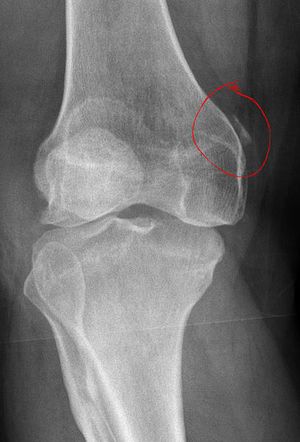
Imaging Studies: PSS is confirmed by using X-rays to find calcification close to the medial femoral condyle.
X-ray showing calcification of proximal MCL (Right Knee)
Physiotherapy Management
At Elite Physiotherapy and Sports Injury Centre, our approach to managing PSS focuses on alleviating pain, restoring function, and preventing recurrence. Our comprehensive treatment plan includes:
Pain management: Using techniques like cryotherapy to lessen discomfort and inflammation.
Manual therapy: Increasing flexibility and decreasing stiffness by mobilizing joints and soft tissues.
Therapeutic Exercises: Creating customized workout plans to improve knee stability by strengthening the hip abductors, hamstrings, and quads.
Advanced Modalities: Incorporating state-of-the-art treatments available at our centre, including:
Shock Wave Therapy: Using acoustic waves to promote tissue healing and lessen pain.
CRET Therapy: Using high-frequency electrical currents, capacitive-resistive electric transfer (CRET) therapy improves tissue healing and lowers inflammation.
SIS: The Super Inductive System (SIS) uses electromagnetic fields to activate muscles and relieve pain by stimulating neuromuscular systems.
High-Intensity Class IV Laser Therapy: Using deep-penetrating laser light, high-intensity class IV laser therapy reduces inflammation and speeds up tissue recovery.
Hydrotherapy: Hydrotherapy uses water-based exercises to increase flexibility and strength while reducing joint tension.
Our holistic approach ensures that each patient receives personalized care tailored to their specific needs, facilitating optimal recovery and return to activity.
Conclusion
Treatment for Pellegrini-Stieda Syndrome must be multimodal. To deliver efficient, customized care, Elite Physiotherapy and Sports Injury Centre combines cutting-edge therapeutic techniques with professional evaluations. Our dedication to using state-of-the-art therapies guarantees our patients the greatest results.

