PLANTAR FASCIITIS

Introduction

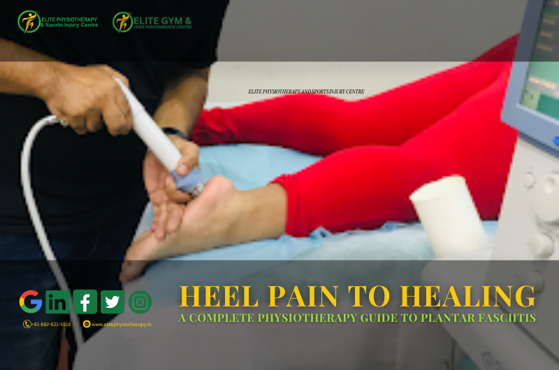
Plantar fasciitis is one of the most common causes of heel pain, affecting athletes and non-athletes alike. Our innovative physiotherapy treatments for plantar fasciitis at Elite Physiotherapy & Sports Injury Centre guarantee a quick and efficient recovery. A thorough explanation of plantar fasciitis, including its causes, symptoms, diagnosis, and state-of-the-art physiotherapy treatment, is given in this article.

What is Plantar Fasciitis?

The inflammation of the plantar fascia, a thick band of tissue that runs from the heel bone to the toes, is known as plantar fasciitis. It is essential for maintaining the arch of the foot and absorbing shock when moving. Pain and stiffness are caused by tiny tears that form when this fascia is overstressed.

Causes of Plantar Fasciitis

Excessive strain and stress on the plantar fascia causes inflammation and microtears, which is known as plantar fasciitis. Typical reasons include:

  • Overuse Injury: Repetitive stress from sprinting, jumping, or extended standing can result in overuse injuries.
  • Poor Foot Biomechanics: High arches, flat feet, or inappropriate running or walking patterns.
  • Inappropriate Footwear: Wearing shoes with insufficient cushioning or arch support is considered inappropriate footwear.
  • Obesity: Excess weight puts undue strain on the plantar fascia.
  • Tight Calf Muscles: The heel is under more stress when ankle mobility is restricted.
  • Sudden Increase in Activity: Increasing training intensity quickly without the necessary conditioning.

Excessive loading of the plantar fascia leads to degenerative changes rather than acute inflammation. Chronic pain and stiffness are caused by microtears that form at the fascia’s attachment to the calcaneus (heel bone). The fascia thickens and fibroses as a result of this tension over time, losing its flexibility and functionality.

Signs, Symptoms, and Clinical Features

  • Heel Pain (First-Step Pain): Severe heel pain, particularly in the morning or after extended periods of inactivity.
  • Localized Tenderness: Palpable pain near the medial calcaneal tubercle.
  • Activity-Aggravated Pain: Prolonged standing, running, or walking exacerbates pain.
  • Foot stiffness: The arch of the foot becomes less flexible, particularly after inactivity.
  • Pain Reduction with Rest: When you take a break, your symptoms go away, but when you move, they come back.
  • Swelling: In rare instances, there may be mild heel edema.

Diagnosis of Plantar Fasciitis at Elite Physiotherapy & Sports Injury Centre

To properly diagnose plantar fasciitis, we at Elite Physiotherapy & Sports Injury Centre perform thorough physical and functional evaluations. We assess foot mechanics, muscular imbalances, and pain sources using specific physiotherapy exams.

Physical and Functional Assessment

1. Observation and Palpation

  • Look for any indications of an irregular weight distribution, foot arch height, and swelling.
  • To determine whether the medial calcaneal tubercle is painful, palpate it.

2. Special Physiotherapy Tests

  • The Windlass Test – Involves dorsiflexing the big toe to assess the tightness of the plantar fascia.
  • The heel raise test – Detects weakness and soreness when performing single-leg heel raises.
  • The Navicular Drop Test – Evaluates arch stability and foot pronation.

3. Gait and Biomechanical Analysis

  • Examining running and walking patterns to identify irregularities in foot action.
  • recognizing compensatory motions that could exacerbate discomfort.

4. Range of Motion and Strength Testing

  • Evaluating calf muscle stiffness, foot arch flexibility, and ankle dorsiflexion.
  • Assessing the strength of the intrinsic foot muscles.

Advanced Physiotherapy Management of Plantar Fasciitis

At Elite Physiotherapy & Sports Injury Centre, we utilize cutting-edge physiotherapy treatments to ensure rapid and effective recovery. Our treatment approach focuses on reducing pain, improving biomechanics, and restoring foot function.

1. Pain Management Techniques

  • Shockwave therapy – Decreases chronic pain, promotes healing, and breaks down scar tissue.
  • High-Intensity Class 4 Laser Therapy – Encourages tissue regeneration and reduces inflammation.
  • Super Inductive System (SIS) – Deep tissue stimulation is provided by the Super Inductive System (SIS) to enhance neuromuscular function and lessen discomfort.
  • Cryotherapy – Cold therapy, or cryotherapy, is used to lessen acute pain and inflammation.

2. Manual Therapy

  • Myofascial Release: Specific manual procedures to alleviate calf muscle and plantar fascia strain.
  • Deep Tissue Massage: Reduces lower leg and foot trigger points.
  • Joint Mobilization: Enhances movement patterns by increasing ankle and foot mobility through joint mobilization.

3. Therapeutic Exercises

Stretching Exercises

  • Plantar Fascia Stretch – Pulling the toes toward the shin to release tension is known as a plantar fascia stretch.
  • Calf Stretching – To lessen the strain on the fascia, stretch the gastrocnemius and soleus muscles.

Strengthening Exercises

  • Intrinsic Foot Muscle Strengthening: Arch lifts, toe curls, and towel scrunches are exercises that strengthen the muscles of the feet.
  • Calf Raises: Increasing the strength of the lower leg and foot muscles.
  • Resisted Ankle Movements: To increase foot stability, use resistance bands.

Balance and Proprioception Training

  • Single Leg Balance Exercise: Improve arch control and foot stability
  • Wobble board training: Training on a wobble board increases foot strength and coordination

4. High-End Electrotherapy and Modalities

  • CRET Therapy (Capacitive-Resistive Energy Transfer Therapy) – Enhances deep tissue healing and reduces inflammation.
  • Hydrotherapy – Water-based exercises to reduce weight-bearing stress on the foot.
  • Ultrasound Therapy – Provides deep tissue healing and improves circulation.

5. Footwear Modification and Orthotics

  • Custom Orthotics – Provides arch support and redistributes pressure evenly.
  • Footwear Advice – Guidance on selecting shoes with proper cushioning and arch support.

6. Kinesiology Taping

  • Supportive Taping Techniques – Reduces stress on the plantar fascia and improves foot mechanics.
  • Dynamic Taping – Provides proprioceptive feedback to enhance foot function.

Why Choose Elite Physiotherapy & Sports Injury Centre for Plantar Fasciitis Treatment?

At Elite Physiotherapy & Sports Injury Centre, we provide:

  • Expert Physiotherapists specializing in sports injury rehabilitation.
  • State-of-the-art technology for advanced pain relief and healing.
  • Personalized treatment plans tailored to individual needs.
  • Holistic approach integrating manual therapy, exercises, and high-end modalities.
  • Fast recovery and long-term pain relief with advanced physiotherapy techniques.

Conclusion

Plantar fasciitis can be debilitating, but with the right physiotherapy treatment, recovery is possible. At Elite Physiotherapy & Sports Injury Centre, we use the latest evidence-based techniques, high-end modalities, and expert physiotherapy care to ensure optimal healing. If you’re experiencing heel pain, don’t let it affect your daily activities. Book an appointment today and take the first step towards a pain-free life!

TURF TOE | TOE INJURY

Introduction

Turf toe is a significant concern for athletes, particularly those engaged in sports requiring sudden acceleration, deceleration, or directional changes. The diagnosis and treatment of turf toe is our specialty at Elite Physiotherapy and Sports Injury Centre, guaranteeing a quick and secure return to optimal performance. ​

Understanding Turf Toe

Turf toe refers to a sprain of the metatarsophalangeal (MTP) joint of the big toe. Usually, this injury happens when the toe is forcefully twisted upward, causing the soft tissues and ligaments around it to stretch or rip. ​

Causes and Mechanism of Injury

The primary cause of turf toe is hyperextension of the big toe, often resulting from:​

  • Sports Activities: Because artificial grass has less traction, playing sports on it might cause the foot to slip backward while the toe stays planted. ​
  • Inappropriate Footwear: The danger might be increased by wearing shoes with flexible soles or inadequate support. ​
  • Repetitive Stress: The disorder may be exacerbated by constant push-off motions, which are typical during running and leaping. ​

Signs and Symptoms

Individuals with turf toe may experience:​

  • Pain and Tenderness are located at the base of the big toe, intensifying during movement.​
  • Swelling and Bruising: Visible around the MTP joint.​
  • Limited Joint Movement: Difficulty in bending or straightening the toe.​
  • Instability: A sensation of looseness in the toe joint.

Diagnostic Methods

At Elite Physiotherapy and Sports Injury Centre, our diagnostic approach includes:​

  • Clinical evaluation: Entails reviewing the patient’s medical history and performing a physical examination to detect limitations in the range of motion, edema, and soreness. ​
  • Functional assessment: Involves tracking weight-bearing and walking patterns to identify any irregularities. ​
  • Special Physiotherapy Tests: Making use of certain evaluations to assess the MTP joint’s and the surrounding structures’ integrity. ​
  • Imaging Studies: Making a referral for MRI or X-rays when required to evaluate soft tissue injury or rule out fractures. ​

Advanced Physiotherapy Management

Our centre offers a comprehensive, individualized treatment plan focusing on:​

Initial Care

  • Rest and Activity Modification: Helping patients cut back on activities that make their symptoms worse. ​
  • Cryotherapy: Using ice packs to relieve pain and inflammation. ​
  • Compression and Elevation: To reduce swelling, apply bandages and raise the foot.

Therapeutic Modalities

  • Shock Wave Therapy: Using acoustic waves to promote healing and reduce pain. ​
  • High-Intensity Class 4 Laser Therapy: This method reduces inflammation and promotes cellular healing by penetrating tissues with laser light. ​
  • Super Inductive System (SIS): Using electromagnetic fields to improve neuromuscular function and reduce pain. ​
  • Hydrotherapy: Performing regulated aquatic activities to increase mobility without putting undue strain on the toe.

Manual Therapy

  • Joint mobilization: It is the process of restoring normal joint mechanics with mild movements. ​
  • Soft Tissue Manipulation: Applying pressure to the surrounding muscles and ligaments to ease stress and increase blood flow.

Exercise Rehabilitation

  • Range of Motion Exercises: Using motions to preserve and improve joint flexibility. ​
  • Strengthening exercises: Targeting the lower leg and foot muscles to improve MTP joint support. ​
  • Proprioceptive Training: Improving balance and coordination through proprioceptive training can help avoid further injuries.

Education and Prevention

  • Advice for Footwear: To safeguard the toe joint, shoes with the right amount of support and stiffness are advised. ​
  • Activity Modification: Advising on methods and approaches to reduce the chance of re-injury when playing sports and going about daily tasks. ​

Conclusion

Although common among athletes, turf toe requires prompt and effective treatment to prevent long-term issues. At Elite Physiotherapy and Sports Injury Centre, we provide customized treatment using cutting-edge modalities and research-proven techniques. We want to ensure that you can quickly and safely resume your active lifestyle.

LISFRANC INJURY OF THE FOOT

Introduction

A Lisfranc injury affects the midfoot, involving the tarsometatarsal joints and associated ligaments. A prompt and precise diagnosis is essential for successful treatment and the best possible outcome. Each patient receives individualized care at Elite Physiotherapy and Sports Injury Centre, where we specialize in cutting-edge physiotherapy therapies designed to address Lisfranc issues. ​

Causes and Mechanism of Injury

Lisfranc injuries can result from both direct and indirect trauma:​

  • Direct Trauma: A force exerted to the top of the foot, as in car accidents, crush injuries, or falls from a height, is frequently the cause of direct trauma. ​
  • Indirect Trauma: Twisting on a plantarly flexed foot or axial loading through the foot are common causes of indirect trauma, which is commonly observed in sports like football or horseback riding. ​

Signs and Symptoms

Patients with a Lisfranc injury may exhibit:​

  • Pain and swelling in the Midfoot.​
  • Bruising on the bottom of the foot.​
  • Pain when bearing weight, particularly when going downstairs.​
  • Tenderness when the midfoot region is touched.​
  • Difficulty or inability to bear weight on the affected foot. ​

Diagnostic Methods

Accurate diagnosis involves a combination of clinical assessment and imaging studies:​

  • Clinical Examination: Key assessments include:​
    • keeping an eye out for bruises and swelling, especially on the bottom of the foot.​
    • Palpating the midfoot to identify areas of tenderness.​
  • Performing specific tests, such as:​
    • Piano-Key Test: Moving the metatarsals up and down to detect pain or instability.
    • Midfoot Compression Test: Squeezing the foot’s width to stress the space between the first and second metatarsals, noting any pain or clicking sounds.
    • Passive Pronation-Abduction Test: Rotating the forefoot outward while stabilizing the hindfoot to assess for pain, indicating potential injury.
  • Imaging Studies:
    • X-Rays: Weight-bearing X-rays are essential to reveal joint misalignments or fractures.​
    • CT Scans: Useful for detecting subtle fractures and planning surgical interventions.​
    • MRI: Effective in evaluating ligament injuries and assessing soft tissue damage. ​

Physiotherapy Management at Elite Physiotherapy and Sports Injury Centre

At Elite Physiotherapy and Sports Injury Centre, we offer a comprehensive physiotherapy program for Lisfranc injuries, focusing on:​

  • Edema Reduction: Utilizing techniques like cryotherapy to minimize swelling and promote healing.​
  • Strengthening Exercises: Addressing muscle weakness resulting from immobilization through targeted exercises.​
    • Toe Flexor and Extensor Strengthening
    • Ankle and Foot Isometric Strengthening
    • Resistance Band Exercises
    • Heel and Arch Strengthening
    • Balance and Proprioception Training
    • Functional and Sport-Specific Strengthening
  • Flexibility Training: Improving range of motion in the foot and ankle to restore standard movement patterns.​
    • Toe and Forefoot Flexibility Exercises
    • Ankle and Midfoot Mobilization Stretches
    • Calf and Achilles Flexibility Exercises
    • Plantar Fascia and Foot Arch Flexibility
    • Functional Dynamic Stretching
  • Gait Training: Assisting patients in regaining proper walking patterns to prevent compensatory injuries.​
  • Custom Orthotics: Providing personalized foot orthoses to support the midfoot and enhance stability during recovery. ​
  • Advanced Modalities Offered
    To enhance recovery, we incorporate state-of-the-art modalities, including:​
    • Shock Wave Therapy: Stimulates healing in damaged tissues and reduces pain.​
    • High-Intensity Class IV Laser Therapy: Penetrates deep tissues to alleviate pain and inflammation.​
    • Hydrotherapy: Utilizes water’s buoyancy to facilitate gentle movement and reduce weight-bearing stress.​
    • Cryotherapy: Cold treatments are applied to decrease inflammation and numb acute pain.​

These advanced treatments are integrated into personalized rehabilitation plans to optimize healing and restore function effectively.

Conclusion

Effective management of a Lisfranc injury requires a thorough understanding of its causes, accurate diagnosis, and a tailored physiotherapy approach. At Elite Physiotherapy and Sports Injury Centre, our dedicated team employs advanced diagnostic techniques and innovative treatment modalities to ensure the best possible outcomes for our patients. If you suspect a Lisfranc injury, seek professional assessment promptly to initiate appropriate care and facilitate a successful recovery.​

SHIN SPLINTS OR MEDIAL TIBIAL STRESS SYNDROME

What is Shin Splints (Medial Tibial Stress Syndrome)?

Shin splints, sometimes referred to as medial tibial stress syndrome (MTSS), are a common overuse injury among athletes, especially runners and those who perform repetitive lower-leg stress-related tasks.

Causes and Mechanism of Injury

Shin splints arise from excessive and repetitive stress on the tibia and surrounding musculature. Key contributing factors include:

  • Training load errors: The tibial bone’s ability to reconstruct and strengthen can be overloaded by abrupt increases in training volume or intensity without sufficient adaption times, which can cause micro-damage.
  • Biomechanical Factors: Overpronation, when the foot rolls inward excessively during walking, can result in an irregular distribution of stress along the shin, raising the risk of MTSS.
  • Muscle Imbalances: Shin pain may result from changes in load distribution caused by weakness or tightness in the calf muscles, especially the soleus.

Signs and Symptoms

Individuals with MTSS typically experience:

  • Diffuse Pain: Aching along the inner edge of the tibia, usually in the lower two-thirds of the leg.
  • Tenderness: The medial part of the shin is sensitive to touch.
  • Activity-Related Discomfort: Pain that worsens when you move and goes away when you relax; however, in more severe cases, the pain may linger even when you’re at rest.

Diagnostic Methods at Elite Physiotherapy and Sports Injury Centre

At Elite Physiotherapy and Sports Injury Centre, our comprehensive assessment for MTSS includes:

  • Clinical Evaluation: To determine pain patterns and contributing variables, a thorough patient history and physical examination are conducted.
  • Functional Assessment: Examining movement and gait patterns to identify anomalies that might be causing shin pain.
  • Special Physiotherapy Tests: Using particular tests to assess biomechanical alignment, muscle strength, and flexibility.

Physiotherapy Management at Elite Physiotherapy and Sports Injury Centre

Our tailored approach to managing MTSS encompasses:

  • Activity Modification: Offering advice on short-term activity reduction or adjustment to ease tibial stress and promote tissue repair.
  • Manual therapy: Reducing muscle tension and increasing flexibility by applying methods like myofascial release and soft tissue mobilization.
  • Therapeutic Modalities: Employing advanced treatments, including:
    • Extracorporeal Shock Wave Therapy (ESWT): By applying shock waves to the injured area, extracorporeal shock wave therapy (ESWT), a non-invasive treatment, encourages recovery.
    • High-Intensity Class IV Laser Therapy: Through deep tissue penetration, high-intensity class IV laser therapy improves tissue healing and lowers inflammation.
    • Cryotherapy: Using cold therapy to lessen inflammation and pain.
    • Hydrotherapy: Water-based workouts to increase strength and flexibility with minimal weight-bearing stress.
  • Strengthening Exercises: To increase load distribution and resilience, create customized training regimens that target the calf muscles, especially the soleus.
  • Stretching regimens: To improve flexibility and lessen tibial strain, use stretching exercises that target the calf muscles.
  • Gait Retraining: Teaching patients how to run properly, including how to change their cadence and foot strike, to reduce undue strain on their shins.
  • Assessment of Footwear: Giving advice on suitable footwear and, if required, custom orthotics to address biomechanical problems such as overpronation. We also provide customized insoles and sandals after a 3D examination of your foot at our clinic.

Conclusion

With a thorough and customized treatment plan, medial tibial stress syndrome is a treatable condition. To guarantee the best possible recovery and a safe return to exercise, we at Elite Physiotherapy and Sports Injury Centre combine cutting-edge therapeutic methods with individualized rehabilitation plans. To start a customized healing journey, speak with our Specialized staff if you’re suffering from shin pain.

Tarsal Tunnel Syndrome

What Is Tarsal Tunnel Syndrome?

A compressive neuropathy known as Tarsal Tunnel Syndrome (TTS) affects the posterior tibial nerve as it travels through the tarsal tunnel, a small opening on the inside of the ankle. This disorder can affect everyday activities and athletic performance by causing pain, tingling, and numbness in the foot and ankle. ​

Causes and Mechanism of Injury

Compression of the posterior tibial nerve within the tarsal tunnel results in TTS. There are several reasons why this compression may occur, such as: ​

  • Anatomical Abnormalities: The nerve may be more stressed by flat feet or fallen arches. ​
  • Trauma: Swelling or scar tissue from ankle sprains or fractures can pinch nerves. ​
  • Systemic Conditions: Inflammation or damage to nerves can be a result of conditions like diabetes or arthritis. ​

For example, An athlete who sprains their ankle may produce scar tissue as the injury heals. TTS might result from this scar tissue encroaching on the tarsal tunnel and squeezing the posterior tibial nerve. ​

Signs and Symptoms

Individuals with TTS may experience:

  • Pain: A burning or aching feeling on the bottom of the foot or inside the ankle. ​
  • Numbness: A “pins and needles” sensation or diminished feeling in the foot. ​
  • Tingling: Prickly feelings that could spread to the toes.
  • Weakness: Feeling unsteady or having trouble moving your feet. ​

Prolonged standing, walking, or physical activity frequently makes symptoms worse; resting may help them.

Diagnostic Methods at Elite Physiotherapy and Sports Injury Centre

At Elite Physiotherapy and Sports Injury Centre, we employ comprehensive physical and functional assessments to diagnose TTS accurately. Our diagnostic approach includes:

  • Clinical examination: Includes measuring muscle strength and reflexes, determining sensitive spots, and assessing foot and ankle alignment. ​
  • Special Physiotherapy Tests: Applying certain tests to elicit symptoms and validate the diagnosis. ​
    • Dorsiflexion-Eversion Test: The therapist extends all toes, everts the foot, and maximally dorsiflexes the ankle. While tapping over the tarsal tube, this posture is maintained for 5–10 seconds. Symptoms along the nerve distribution are replicated in a positive test. The Student Physical Therapist’s website
    • Tinel’s Sign: A “pins and needles” or tingling feeling in the foot caused by light tapping over the tarsal tunnel indicates nerve irritation. ​

Physiotherapy Management at Elite Physiotherapy and Sports Injury Centre

Our center offers a multifaceted approach to managing TTS, focusing on alleviating symptoms, addressing underlying causes, and preventing recurrence. Our advanced physiotherapy treatments include:

  • Manual therapy: Methods to improve mobility and lessen nerve compression, such as soft tissue manipulation and nerve gliding exercises. ​
  • Therapeutic Exercises: Tailored stretching and strengthening regimens that focus on the ankle and foot muscles to enhance function and support. ​
  • Orthotic Intervention: Reducing tension on the posterior tibial nerve by correcting biomechanical anomalies such as flat feet with custom-made orthotics.
  • High-End Modalities: Using cutting-edge therapies to hasten recovery and lessen symptoms ​
    • Shock Wave Therapy: Reduces pain and encourages tissue regeneration by using acoustic waves. ​
    • CRET Therapy: Radiofrequency energy is used in Capacitive Resistive Electric Transfer (CRET) therapy to promote deep tissue repair and decrease inflammation. ​
    • SIS: By stimulating neuromuscular areas with high-intensity electromagnetic fields, the Super Inductive System (SIS) reduces pain and speeds up the healing process. ​
    • High-Intensity Class IV Laser Therapy: Reduces inflammation and speeds up tissue repair by penetrating deep tissues with concentrated laser energy. ​
    • Hydrotherapy: In order to lessen the strain on the injured area while preserving mobility, hydrotherapy involves patients in water-based exercises. ​
    • Cryotherapy: Reduces inflammation and numbs acute pain by applying regulated cold exposure. ​

At Elite Physiotherapy and Sports Injury Centre, our skilled physiotherapists are committed to offering individualized treatment. We customize each treatment plan to meet the unique demands of each patient, guaranteeing a full recovery and a return to sports and daily activities. ​

Make an appointment at Elite Physiotherapy and Sports Injury Centre if you suffer from Tarsal Tunnel Syndrome symptoms. Our team provides compassionate care and state-of-the-art treatments to support your recovery journey. ​

HAGLUND’S DEFORMITY

What is Haglund’s deformity?

A bony growth on the rear of the heel, where the Achilles tendon connects to the calcaneus (heel bone), is known as Haglund’s deformity. This illness frequently causes pain, swelling, and discomfort, which majorly impacts day-to-day functioning and sports performance. Our specialty at Elite Physiotherapy and Sports Injury Centre is the thorough evaluation and treatment of Haglund’s deformity through the use of state-of-the-art physiotherapy methods and innovative treatment approaches.

Causes and Mechanism of Injury

Repetitive pressure, friction, and biomechanical abnormalities aggravating the soft tissues surrounding the heel bone cause Haglund’s deformity. Its development is influenced by several factors, including:

  • Genetic Predisposition: Excessive pressure on the rear of the heel is more likely to occur in people with tight Achilles tendons or high arches in their feet.
  • Inappropriate Footwear: The issue may worsen if you wear tight, stiff, or high-heeled shoes that cause friction against the heel bone.
  • Overuse and Repetitive Stress: The Achilles tendon and heel bone are constantly under stress from athletes and participants in sports like running, sprinting, and jumping.
  • Poor Biomechanics: Excessive pronation, excessive supination, or abnormal walking patterns can all lead to excessive pressure on the heel’s back.
  • Tight Achilles Tendon: A shortened Achilles tendon can cause limited ankle dorsiflexion, which can put more strain on the heel bone.

Signs and Symptoms of Haglund’s Deformity

The common symptoms include:

  • There is a noticeable bony mass on the heel’s back.
  • Pain and tenderness at the enlargement location, particularly when running or walking.
  • Swelling and redness are brought on by inflammation of the soft tissues around it.
  • Achilles tendon inflammation causes stiffness and restricted ankle movement.
  • Increased pain when wearing shoes, particularly ones with stiff heel counters.

Diagnostic Methods at Elite Physiotherapy and Sports Injury Centre

For assessing the degree and impact of Haglund’s deformity, we at Elite Physiotherapy and Sports Injury Centre use a comprehensive diagnostic process that consists of both functional and physical evaluations.

Clinical and Functional Assessment

  • Visual Inspection: Recognizing the distinctive heel bump, redness, and swelling.
  • Palpation: Measuring localized swelling and discomfort to gauge the level of irritation.
  • Range of Motion (ROM) Tests: Assessing Achilles tendon and ankle flexibility to identify motion restrictions.
  • Gait analysis is the process of evaluating walking patterns in order to find biomechanical anomalies that are causing excessive heel stress.
  • Special Physiotherapy Tests:
    • Passive Ankle Dorsiflexion Test: To determine how tight the Achilles tendon is, use the passive ankle dorsiflexion test.
    • Heel Raise Test: The heel raise test is used to assess mobility and discomfort when using the calf muscles.
    • Tibial Nerve Stretch Test: To rule out nerve involvement causing posterior heel discomfort, use the tibial nerve stretch test.

Physiotherapy Management at Elite Physiotherapy and Sports Injury Centre

In order to prevent recurrence, our physiotherapy-based treatment aims to improve mobility, lessen pain, and address the underlying biomechanical problems.

1. Management of Pain and Inflammation

  • Cryotherapy: Often known as ice therapy, numbs severe pain and reduces swelling.
  • Shock Wave Therapy: By improving blood circulation, shock wave therapy promotes tissue healing and lowers inflammation.
  • High-Intensity Class 4 Laser Therapy: Provides deep-penetrating light energy to the afflicted area, accelerating cellular repair.

2. Improving Mobility and Flexibility

  • Manual therapy: mobilization methods to enhance Achilles tendon flexibility and joint mechanics.
  • Stretching Exercises:
    • Stretches for the calf and Achilles to increase range of motion and reduce stress.
    • Stretching the plantar fascia can improve foot mobility.

3. Strengthening the Lower Limb and Foot Muscles

  • Specific Strengthening Exercises:
    • To strengthen and lessen tension on the Achilles tendon, use eccentric heel drops.
    • Exercises for the foot’s intrinsic muscles to improve arch stability.
    • Exercises using resistance bands to stabilize the ankles.

4. Gait training and biomechanical adjustments

  • Super Inductive System: Enhances gait function by activating neuromuscular areas using electromagnetic fields.
  • Custom Orthotics and Footwear Modifications: The purpose of custom orthotics and footwear modifications is to reduce pressure on the heel and correct aberrant biomechanics.
  • Taping and Bracing: Reduces tension in the afflicted area and offers structural support.

5. Cutting-edge techniques for Quicker Recovery

  • CRET Therapy: Promotes blood circulation and deep tissue healing.
  • Hydrotherapy (Aquatic Therapy): By lowering weight-bearing stress, hydrotherapy (also known as aquatic treatment) enables pain-free movement and strengthening.

Conclusion

At Elite Physiotherapy and Sports Injury Centre, we provide state-of-the-art treatments in conjunction with skilled physiotherapy techniques to ensure optimal healing. If you are dealing with chronic heel pain, get in touch with us today for a thorough evaluation and a customized rehabilitation program. Haglund’s deformity can have a significant impact on mobility and overall quality of life, but effective relief and recovery are achievable with an accurate diagnosis and an individualized physiotherapy plan.

Retrocalcaneal Bursitis

What is Retrocalcaneal Bursitis?

The painful inflammatory disease known as retrocalcaneal bursitis affects the bursa between the calcaneus (heel bone) and the Achilles tendon. Athletes and those who perform repetitive tasks that impose excessive tension on the heel are frequently affected by this condition. It can seriously affect everyday activities and mobility if treatment is not received. At Elite Physiotherapy and Sports Injury Centre, we use advanced physiotherapy methods and luxury modalities to diagnose and treat retrocalcaneal bursitis in a comprehensive and sophisticated manner.

Causes and Mechanism of Injury

Repetitive friction and severe pressure on the bursa at the rear of the heel can cause retrocalcaneal bursitis. Among the most common causes are:

  • Overuse and Repetitive Stress: The bursa may get irritated by high-impact exercises including jogging, jumping, and abrupt direction changes.
  • Inappropriate Footwear: Irritation and inflammation can result from wearing stiff or poorly fitting shoes that rub on the heel.
  • Biomechanical Abnormalities: Extreme strain on the Achilles tendon and bursa can result from conditions such as high arches, flat feet, or a changed walking pattern.
  • Trauma or Direct Impact: Inflammation may be brought on by a forceful impact to the rear of the heel.
  • Underlying Medical Conditions: People who have gout, rheumatoid arthritis, or infections are more likely to develop bursitis.

Signs, Symptoms, and Clinical Features

The following symptoms are frequently seen in people with retrocalcaneal bursitis:

  • Pain and tenderness at the back of the heel, especially when walking or doing vigorous activities.
  • Swelling, warmth, and redness, in the affected region.
  • Increased pain when pushing off the foot or standing on toes.
  • Ankle stiffness and limited range of motion, especially in the morning or after extended periods of inactivity.
  • Dorsiflexion, or pulling the toes toward the shin, exacerbates the pain.

Diagnosis at Elite Physiotherapy and Sports Injury Centre

To establish the diagnosis and rule out other possible disorders like Achilles tendinitis or Haglund’s deformity, our center conducts a comprehensive physical and functional assessment. Our diagnostic methodology consists of:

Clinical Assessment

  • Palpation and Inspection: Examining the afflicted bursa for soreness, redness, and swelling.
  • Assessing the ankle joint’s range of motion (ROM): This involves determining how flexible and mobile it is.
  • Strength testing: involves determining how strong the Achilles tendon and calf muscles are.

Functional and Special Physiotherapy Tests

We employ specific physiotherapy tests, such as the following, to validate the diagnosis:

  • Hoffa’s Test: To determine whether Achilles tendon involvement and underlying soft tissue inflammation are present.
  • Arc Sign Test: Distinguishing between bursitis and Achilles tendinopathy.
  • Royal London Hospital Test: Assessing Achilles pain and swelling with the Royal London Hospital Test.
  • The Silfverskiold Test: Measures tension in the gastrocnemius muscles, which can put too much strain on the bursa.

In certain situations, for a more thorough evaluation, we could suggest imaging tests like MRI or ultrasound.

Physiotherapy Management at Elite Physiotherapy and Sports Injury Centre

At our clinic, we treat retrocalcaneal bursitis using customized, evidence-based physiotherapy plans. Our course of treatment consists of:

1. Management of Pain and Inflammation

  • Cryotherapy: Using ice packs to minimize swelling and give instant pain relief is known as cryotherapy.
  • Activity Modification: To avoid more aggravation, it is advised to temporarily limit high-impact activities.

2. Advanced Physiotherapy Modalities

We use state-of-the-art techniques to maximize healing and accelerate recovery, such as:

  • Shock Wave Therapy: Shock wave therapy uses acoustic waves to encourage tissue repair and alleviate pain.
  • CRET Therapy: Promotes inflammation reduction and cellular healing.
  • The Super superinductive system (SIS): Uses electromagnetic fields to promote healing, increase circulation, and reduce pain.
  • High-Intensity Class 4 Laser Therapy: Enhances tissue repair, lessens pain, and speeds up cellular healing.
  • Hydrotherapy: Hydrotherapy includes water-based exercises that increase joint mobility while reducing strain on the injured area.

3. Techniques for Soft Tissue and Manual Therapy

  • Myofascial Release: Myofascial release relieves tension in the Achilles tendon and surrounding structures.
  • Deep Tissue Mobilization: Improves blood flow and eases tense muscles.
  • Joint mobilization: Enhances ankle function and flexibility.

4. Exercises for Therapy

We create specialized workout regimens to regain strength, flexibility, and appropriate biomechanics, such as:

  • Stretching exercises: They target the Achilles tendon and calf muscles to release stress.
  • Strengthening exercises: To increase stability and lessen bursa strain, focus on the calf, ankle, and foot muscles.
  • Proprioception Training: Proprioception training includes exercises for balance and coordination that improve functional mobility and reduce the risk of re-injury.

5. Postural and Biomechanical Adjustment

  • Gait Training: Gait training corrects faulty walking habits to lessen undue heel strain.
  • Orthotic Recommendations: Providing personalized foot insoles for better pressure distribution and shock absorption is one orthotic recommendation.
  • Advice on Footwear: Suggest suitable footwear with supportive arch support and cushioned heels.

Prevention and Long-Term Care

To lower the chance of recurrence, we at Elite Physiotherapy and Sports Injury Centre concentrate on both prevention and treatment methods. We instruct our patients on:

  • Appropriate warm-up and cool-down techniques both before and following physical activity.
  • Maintaining the lower extremities’ strength and flexibility.
  • Gradually increase activity levels to prevent overuse injuries.
  • Maintaining appropriate foot alignment requires the use of orthotics and supportive footwear.

Conclusion

If left untreated, retrocalcaneal bursitis can become incapacitating. At Elite Physiotherapy and Sports Injury Centre, we combine cutting-edge physiotherapy methods, upscale modalities, and customized rehabilitation programs to offer a thorough, patient-centered approach to treatment. Our objectives are to guarantee a full recovery, avoid recurrence, and assist our patients in getting back to their regular activities and sports without any restrictions.

For an extensive evaluation and top-notch physiotherapy treatment, come see us right now if you have heel pain and think you may have retrocalcaneal bursitis.

Achilles Tendinopathy: Cause, Symptoms & Physiotherapy Management

Understanding Achilles Tendinopathy

A common overuse ailment that affects the Achilles tendon, which joins the calf muscles to the heel bone, is Achilles tendinopathy. Pain and stiffness in the back of the heel are common symptoms of this illness, especially in the morning or during exercise. Achilles tendinopathy is our specialty at Elite Physiotherapy and Sports Injury Centre, and we guarantee a customized treatment plan for every patient.

Causes and Mechanism of Injury

Repetitive strain on the tendon without sufficient recovery is the main cause of Achilles tendinopathy. Unexpected increases in physical activity, inappropriate footwear, and biomechanical problems like overpronation are some of the factors that contribute to this illness. For example, an athlete may develop tendinopathy if they suddenly increase their exercise volume without giving their muscles enough time to recover.

Signs and Symptoms

Patients typically report:

  • Achilles tendon pain and stiffness, often following periods of inactivity.
  • Swelling or thickening of the tendon.
  • Discomfort when running or climbing stairs—activities that put stress on the tendon.

Diagnostic Methods at Our Clinic

To diagnose Achilles tendinopathy, we at Elite Physiotherapy and Sports Injury Centre use thorough physical and functional evaluations. Our assessment consists of:

  • Palpation: To find areas of discomfort or thickness, gently press along the tendon.
  • Range of Motion Tests: Measuring ankle flexibility to identify restrictions.
  • Strength testing: Assessing the strength of the calf muscles to find imbalances.
  • Special Physiotherapy examinations: To distinguish Achilles tendinopathy from other disorders, we use specific examinations.

Physiotherapy Management

Achilles tendinopathy is treated in our clinic utilizing a multimodal approach that emphasizes functional restoration, tendon repair, and pain management. Among our methods of treatment are:

  • Eccentric Loading Exercises: These exercises stretch the strained tendon while encouraging collagen realignment and strengthening.
  • Shock Wave Therapy: Using acoustic waves to promote healing and lessen pain is known as shock wave therapy.
  • CRET Therapy: By using electromagnetic energy to promote deep heat, Capacitive Resistive Energy Transfer (CRET) therapy improves tissue recovery.
  • Super Inductive System: High-intensity electromagnetic fields are used in the Super Inductive System to reduce pain and promote tissue regeneration.
  • High-Intensity Class 4 Laser Therapy: High-intensity Class 4 laser therapy uses deep tissue laser treatments to lower inflammation and accelerate healing.
  • Hydrotherapy: Performing exercises in warm water to increase flexibility and strength while reducing tendon stress.
  • Cryotherapy: Cryotherapy uses cold therapies to reduce inflammation and dull severe pain.

Our skilled physiotherapists create customized treatment programs to guarantee that every patient gets the best care possible based on their unique requirements.

Strategies for Prevention

In order to avoid recurrence, we recommend:

  • Increase activity levels gradually to prevent overtaxing the tendon.
  • Wear supportive shoes that suit the situation.
  • Including consistent calf muscle strengthening and stretching workouts.

Our goal at Elite Physiotherapy and Sports Injury Centre is to help patients recover quickly and effectively by offering cutting-edge, research-based therapies for Achilles tendinopathy.

Calcaneal Fractures: Causes, Symptoms, and Advanced Physiotherapy Management

Calcaneal Fractures

The calcaneus bones in the foot are injured in calcaneal fractures. The mobility and quality of life might be greatly affected by these fractures. Our specialty at Elite Physiotherapy and Sports Injury Centre is thorough rehabilitation for these injuries, using cutting-edge techniques to guarantee the best possible outcome.

Causes and Mechanism of Injury

Axial force is usually the cause of calcaneal fractures, which are the most frequent tarsal bone fractures. This occurs when someone falls from a height and lands on their feet, compressing and breaking the heel bone. If not properly treated, such injuries can result in serious morbidity.

Signs and Symptoms

Individuals who have calcaneal fractures may suffer from:

  • Severe hindfoot pain.
  • Bruises and swelling near the heel or ankle.
  • Unable to use the injured foot to support the weight.
  • Pain or deformity in the ankle or heel area.

Diagnostic Methods

Our assessment at Elite Physiotherapy and Sports Injury Centre consists of the following:

  • Physical Examination: Evaluating edema, bruising, and deformity.
  • Functional Assessment: Evaluating weight-bearing ability and range of motion.
  • Special Physiotherapy Tests: Using certain tests to detect ligamentous injuries and fractures.

Advanced Physiotherapy Management

Our goal when treating calcaneal fractures is to minimize pain, restore function, and avoid complications. To meet the needs of each patient, we combine modern techniques, therapeutic exercises, and manual treatment.

High-End Modalities Utilized

We use the following cutting-edge therapies to improve recovery:

  • Shock Wave Therapy: Applying acoustic waves to the wounded area helps to promote tissue repair and lessen discomfort.
  • CRET Therapy: Through the use of electromagnetic energy, Capacitive Resistive Energy Transfer (CRET) Therapy improves deep tissue healing while lowering inflammation and increasing circulation.
  • The Super Inductive System:  Stimulates muscles and speeds up bone mending by using strong electromagnetic fields.
  • High-Intensity Class 4 Laser Therapy: Promotes quicker tissue repair by reducing pain and inflammation with concentrated light energy.
  • Hydrotherapy: Uses water-based workouts to reduce joint tension and increase strength and mobility.
  • Cryotherapy: In the early stages of an injury, cryotherapy uses cold therapy to minimize discomfort and swelling.

Personalized Rehabilitation Programs

Each patient receives a personalized rehabilitation program from us that emphasizes:

  • Pain management: is the process of reducing discomfort by using physical techniques and modalities.
  • Restoring Range of Motion: To restore mobility, use stretching techniques and joint mobilizations.
  • Strengthening exercises: To regain strength and function, focus on the lower leg and foot muscles.
  • Gait training: Gait training helps patients rediscover appropriate walking techniques to avoid further injuries.
  • Proprioceptive training: Using certain exercises to improve balance and coordination.

Our goal at Elite Physiotherapy and Sports Injury Centre is to treat calcaneal fractures with state-of-the-art physiotherapy techniques. We design our individualized care programs and cutting-edge modalities to promote a quick and efficient recovery, allowing our patients to confidently resume their regular activities.

Anterior Ankle Tendinopathy: Diagnosis & Advanced Physiotherapy Management

Anterior Ankle Tendinopathy

Pain and dysfunction at the front of the ankle are symptoms of anterior ankle tendinopathy, which mostly affects the tibialis anterior tendon. Both daily activities and athletic performance may be greatly impacted by this condition, particularly for those who do repetitive dorsiflexion movements.

Causes and Mechanism of Injury

To dorsiflex the foot and enable upward movement, the tibialis anterior muscle is essential. Microtrauma and tendon degeneration can result from overuse or repetitive strain, especially during activities like jogging or climbing on uneven terrain. People can be predisposed to this tendinopathy by systemic disorders like diabetes or hypertension, as well as intrinsic variables including age-related tendon degradation and diminished vascularity. Mechanical overload, abrupt increases in exercise intensity, and inappropriate footwear are examples of extrinsic variables.

Long-distance runners who abruptly increase their training volume without enough recovery frequently develop anterior ankle tendinopathy. Running-induced repetitive dorsiflexion and inadequate recovery time might overstress the tibialis anterior tendon, causing microtrauma and subsequent tendinopathy.

Signs and Symptoms

Patients typically present with:

  • Pain in the medial midfoot and anterior ankle.
  • Resistance to dorsiflexion worsens the pain.
  • Tenderness along the tibialis anterior tendon’s length.
  • Crepitus or swelling in the affected area.

These symptoms may be more noticeable at night and frequently get worse with activity.

Diagnostic Methods at Elite Physiotherapy and Sports Injury Centre

We at Elite Physiotherapy and Sports Injury Centre use thorough physical and functional evaluations to diagnose anterior ankle tendinopathy. Our strategy consists of:

  • Clinical Evaluation: a thorough medical history to determine the development of symptoms, activity levels, and possible causes.
  • Physical Examination: Palpation along the tibialis anterior tendon to detect tenderness or edema; discomfort assessment using resisted dorsiflexion.
  • Special Tests: Orthopedic specialists use particular tests to assess the integrity and function of tendons.

These evaluations enable us to precisely identify the condition and provide a customized therapy strategy.

Physiotherapy Management at Elite Physiotherapy and Sports Injury Centre

In order to prevent recurrence, our facility provides a thorough physiotherapy program that addresses underlying reasons in addition to symptom relief. Among our managerial techniques are:

  • Activity Modification: Helping patients cut back on or stop activities that make their symptoms worse so that the tendon can recover.
  • Manual therapy: Using methods like massage and soft tissue mobilization to ease tense muscles and increase blood flow to the afflicted area.
  • Therapeutic Exercises: Creating customized workout plans to improve the tibialis anterior muscle and its surrounding structures’ strength, flexibility, and endurance.
  • High-end modalities: Making use of cutting-edge therapeutic technologies to ease pain and encourage recovery, such as:
  1. Shock Wave Therapy: Shock wave therapy involves using acoustic waves to promote tendon repair and reduce pain.
  2. Capacitive Resistive Electric Transfer (CRET) Therapy: Using deep thermal effects, Capacitive Resistive Electric Transfer (CRET) therapy improves tissue repair.
  3. Super Inductive System: The Super Inductive System uses high-intensity electromagnetic fields to lessen pain and promote tissue regeneration.
  4. High-Intensity Class 4 Laser Therapy: High-intensity Class 4 laser therapy involves using deep tissue laser therapy to lower inflammation and encourage healing.
  5. Hydrotherapy: Hydrotherapy involves using water-based exercises to increase strength and mobility while reducing joint stress.
  6. Cryotherapy: Using cold therapy to alleviate acute pain and inflammation.

Pain and dysfunction at the front of the ankle are symptoms of anterior ankle tendinopathy, which mostly affects the tibialis anterior tendon.

At Elite Physiotherapy and Sports Injury Centre, we’re dedicated to giving our patients individualized care and state-of-the-art treatments so they may reach their performance and wellness objectives.