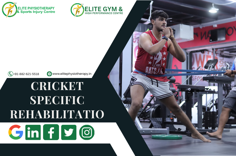
TR-Therapy

Revolutionizing Rehabilitation with TR-Therapy

At Elite Physiotherapy and Sports Injury Centre, we take pride in continuing to lead the way in medical innovation by providing innovative treatments that transform recovery and rehabilitation. One of the most cutting-edge techniques we use is TR-Therapy, a novel therapy designed to speed up recovery, lessen discomfort, and increase general mobility. Modern technology and individualized treatment are flawlessly combined in this therapy, guaranteeing each patient receives the best possible outcomes.

What is TR-Therapy?

Targeted Radiofrequency Therapy, or TR-Therapy for short, is a non-invasive therapeutic approach that uses high-frequency electromagnetic energy to encourage deep tissue repair. In contrast to conventional treatments, TR-Therapy induces regulated hyperthermia (deep heating) by penetrating deeply into muscles, ligaments, and connective tissues. Significant pain reduction, enhanced circulation, and a quicker recovery are all made possible by this special mechanism. The versatility of TR-Therapy, which can be utilized with or without thermal effects, makes it ideal for a variety of injuries and medical disorders.

How TR-Therapy Works?

A closer look into TR-Therapy’s process reveals that it uses a combination of ergonomic applicators and sophisticated radiofrequency radiation to give targeted treatment:

  1. Capacitive Mode: This mode concentrates on soft tissues and the skin, targeting superficial tissues. It’s perfect for ailments like small sprains or inflammations that need to be treated at the surface level.
  2. Resistive Mode: The resistive mode targets deeper structures, such as tendons, joints, and muscles. It works especially well for joint stiffness, deep muscle injuries, and persistent pain.
  3. Dynamic Impedance Control: During therapy, a state-of-the-art function called Dynamic Impedance Control continuously analyzes tissue impedance. This guarantees the best possible energy delivery, modifying in real time for optimal efficacy and comfort.

Patients frequently report feeling a calming warmth that eases tension and relaxes muscles during therapy. TR-Therapy’s combination of thermal and physical impacts makes it both fun and effective.

Applications of TR-Therapy at Elite Physiotherapy and Sports Injury Centre

At our center, we include TR-Therapy into all-encompassing treatment programs to address a variety of ailments. Here are a few of the most important uses:

1. Management of Chronic Pain

The quality of life can be greatly impacted by chronic pain, particularly in regions like the shoulders, neck, or lower back. Deep tissue relaxation, a reduction in muscle tension, and long-term pain relief are all benefits of TR-Therapy. For people with ailments like frozen shoulder, cervical spondylosis, or lumbar pain, this makes it a great choice.

2. Improved Muscle Regeneration

Active people and athletes frequently have small tears, overuse injuries, and muscular weariness. TR-Therapy increases oxygen delivery to tissues and circulation, which speeds up muscle recovery. This therapy helps patients return to optimum performance more quickly, whether they are recovering from strenuous training sessions or treating sports-related injuries.

3. Edema and Post-Traumatic Swelling

Edema and swelling can impede the healing process for those recovering from surgery or trauma. The non-thermal application of TR-Therapy efficiently lowers inflammation and encourages lymphatic drainage, which speeds up healing and increases mobility.

4. Myofascial Pain Syndrome

Widespread pain and stiffness can be caused by trigger points in muscles and fascia. TR-Therapy precisely targets these regions, relieving tension and reestablishing regular muscular function. This is particularly helpful for ailments like tennis elbow, IT band dysfunction, and plantar fasciitis.

5. Tendon and Joint Rehabilitation

Rest and rehabilitation must frequently be carefully balanced in cases of joint and tendon injuries. The focused approach of TR-Therapy guarantees efficient therapy without overtaxing the afflicted area. Because of this, it is the preferred treatment for tendinitis, ligament sprains, and arthritis.

Why Choose Elite Physiotherapy and Sports Injury Centre?

Elite Physiotherapy & Sports Injury Centre was founded in 2008 and has been at the forefront of revolutionizing rehabilitation and physiotherapy in India. What sets us apart is this:

  • Holistic Rehabilitation: Our method helps patients get back to their regular activities or sports after receiving therapeutic treatment. We place a strong emphasis on result-oriented healing, making sure that patients not only recover but also perform at their best.
  • Cutting Edge Facilities: We provide our patients with the newest developments in medical technology thanks to our state-of-the-art physiotherapy equipment, which includes TR-Therapy systems.
  • Tailored Care Plans: Each patient at Elite Physiotherapy is given a customized treatment plan based on their unique requirements, objectives, and recovery schedules. To optimize benefits, TR-Therapy is smoothly included into these plans.
  • Sports Physiotherapy Expertise: We have years of expertise treating athletes and active people, and our focus is on improving athletic performance and treating sports injuries.

Experience the Difference

At Elite Physiotherapy and Sports Injury Centre, we combine knowledge, technology, and compassion to provide unmatched care. TR-Therapy is evidence of our dedication to quality and innovation in rehabilitation. TR-Therapy can be extremely beneficial whether you’re trying to improve your athletic performance, manage a chronic illness, or recover from an injury.

Stop by our office today to learn more about how you can include TR-Therapy in your healing process. Allow us to assist you in starting along the path to an active and pain-free life.

Cryotherapy

Cryotherapy: A Modern Approach to Recovery and Healing at Elite Physiotherapy and Sports Injury Centre

We at Elite Physiotherapy and Sports Injury Centre are committed to providing cutting-edge therapies to improve sports performance, manage pain, and speed up recovery. Cryotherapy, a ground-breaking method of wellness and rehabilitation, is one of our cutting-edge treatments. The benefits and science of cryotherapy, including the use of cryoair and cryobaths, are discussed in this article along with how these therapies are included into our treatment plans.

What is Cryotherapy?

Cold treatment, also known as cryotherapy, uses extremely low temperatures to treat patients. This technique has been used for many years to lessen pain, promote healing, and reduce inflammation. By narrowing blood vessels and lowering nerve activity in the afflicted location, cryotherapy helps to reduce swelling and relieve pain.

Cryo baths and cryo air are the two main types of cryotherapy that we use at Elite Physiotherapy. Both approaches work well, and how they are used will rely on the patient’s particular requirements.

Cryo Bath: Submersion Therapy for Full-Body Recovery

Cryo baths, also called ice baths, involve submerging the body (or specific body parts) in cold water, typically maintained between 10°C and 15°C. Many athletes and injury-recovering patients employ this type of hydrotherapy.

Benefits of Cryo Baths:

  1. Reduces Muscle Soreness: After training or competition, athletes frequently choose cryo baths because they are a very effective way to relieve delayed onset muscle soreness (DOMS).
  2. Improves Circulation: The cold water makes blood vessels contract; these vessels then expand as the body temperature returns to normal, increasing blood flow and eliminating waste products from metabolism.
  3. Reduces Inflammation: Cryo baths hasten the healing process by lowering swelling in strained or injured muscles.
  4. Enhances Mental Recovery: The shock of cold water triggers the release of endorphins, elevates mood, and lowers stress levels.

At Elite Physiotherapy, we provide personalized cryo bath treatments to guarantee each patient’s utmost comfort and efficiency. Our cryo baths fulfill your specific needs, whether you are recovering from intense physical activity or a sports injury.

Cryo Air: Targeted Cooling for Precision Therapy

Cryoair therapy targets specific body parts by delivering cooled, dry air through specialized equipment. Without the requirement for submersion in water, the air temperature can drop as low as -30°C to -60°C, producing a powerful cooling effect.

Benefits of Cryo Air Therapy:

  1. Localized Treatment: For sprains, tendinitis, or joint pain, cryo air is perfect for focusing on certain wounds or trouble spots.
  2. Comfortable and Non-Invasive: Patients who are sensitive to cold or wet circumstances may find cryo-air therapy to be a comfortable alternative to ice packs or cryo baths because it does not require direct contact with water.
  3. Fast Recovery: Usually lasting five to fifteen minutes, sessions provide quick results.
  4. Enhances Skin and Tissue Health: The deep cooling promotes the formation of collagen and increases skin suppleness, which is advantageous for the healing process following surgery.

Our cutting-edge cryo-air equipment at Elite Physiotherapy enables accurate temperature control and application, guaranteeing that patients receive the best care possible for their conditions.

Why Choose Cryotherapy at Elite Physiotherapy?

At Elite Physiotherapy and Sports Injury Centre, we provide unmatched care by fusing state-of-the-art technology with our extensive expertise. The purpose of our cryotherapy treatments is to:

  • Accelerate Recovery: Cryotherapy can help you get back to your activities more quickly, whether you’re an athlete or someone who has chronic pain.
  • Enhance Performance: Frequent cryotherapy procedures can maximize general physical performance, decrease weariness, and increase circulation.
  • Personalized Care: After evaluating each patient’s health, we suggest the best cryotherapy technique, whether cryo air, cryo bath, or a combination of the two.

What to Expect During Your Session

Our skilled specialists will walk you through each step of the cryotherapy procedure when you visit Elite Physiotherapy. We make sure the water temperature for cryo baths is both comfortable and efficient, and we modify the time to fit your demands and tolerance. Our professionals apply focused relief using cold air therapy by concentrating on the afflicted area using handheld instruments.

Conclusion

In the fields of sports medicine and rehabilitation, cryotherapy is revolutionary. Elite Physiotherapy and Sports Injury Centre proudly provides this cutting-edge treatment to our patients. Cryotherapy can be a potent complement to your treatment strategy, whether you’re trying to manage chronic pain, recover from an accident, or enhance your athletic performance.

Make an appointment for cryotherapy with us right now to discover the life-changing advantages of cold therapy. Allow us to provide you with the knowledge and attention you need to reach your performance and recovery goals.

Super Inductive System (SIS)

Super Inductive System (SIS): Revolutionizing Physiotherapy at Elite Physiotherapy and Sports Injury Centre

New technologies are changing how injuries are handled and treated in the dynamic field of physiotherapy. The Super Inductive System (SIS), a cutting-edge therapeutic device that blends cutting-edge technology with tried-and-true medical principles to provide unmatched results, is a ground-breaking development. We at Elite Physiotherapy and Sports Injury Centre are proud of being at the forefront of contemporary physiotherapy procedures, and integrating SIS into our practice demonstrates our dedication to giving our patients the finest care possible.

What is the Super Inductive System (SIS)?

The Super Inductive System (SIS) is a noninvasive technology with a wide range of applications that activates neuromuscular structures, improves circulation, and promotes cellular repair. It is an indispensable tool for treating a variety of musculoskeletal conditions. SIS is a revolutionary therapeutic device that uses high-intensity electromagnetic fields to stimulate deep tissue healing and pain relief.

Key Features of SIS:

  1. High-frequency electromagnetic fields: SIS can precisely target tissues since it operates at frequencies between 1 Hz and 150 Hz.
  2. Painless and Non-Invasive: The treatment is completely non-invasive, guaranteeing patient security and comfort.
  3. Versatility: From severe injuries to persistent pain, SIS can treat a wide range of ailments.
  4. Quick Results: After just a few sessions, many patients see a significant improvement.
  5. Customizable Treatments: The system enables therapists to modify treatment plans following each patient’s requirements.

How SIS Works

High-intensity electromagnetic pulses that activate nerve terminals and cause deep muscular spasms are the basis of SIS. These electromagnetic fields are:

  • Boost Blood Circulation: Increased blood flow lowers inflammation and improves tissue healing.
  • Encourage Pain Relief: By interfering with pain signals, electromagnetic waves offer both short-term and long-term pain relief.
  • Promote Bone Healing: SIS is useful in treating osteoporosis and fractures because it promotes osteogenesis.
  • Strengthen Muscles: SIS helps strengthen weak muscles by causing involuntary contractions.

Applications of SIS in Physiotherapy

Elite Physiotherapy and Sports Injury Centre uses The Super Inductive System to treat a variety of ailments, such as:

  1. Chronic Pain: For ailments including fibromyalgia, arthritis, and back pain, SIS offers substantial relief.
  2. Sports Injuries: SIS improves performance and speeds up recovery from sprains, strains, and ligament tears.
  3. Neurological Disorders: The neuromuscular stimulation that SIS provides is beneficial for conditions including paralysis, neuropathy, and spasticity.
  4. Post-Surgical Rehabilitation: SIS reduces the production of scar tissue and promotes a quicker recovery.
  5. Osteoporosis and Bone Fractures: The system is perfect for boosting skeletal health because of its bone-healing capabilities.

Benefits of SIS at Elite Physiotherapy and Sports Injury Centre

To get better results, we at Elite Physiotherapy and Sports Injury Centre have included SIS into our treatment plans. For our patients, SIS is revolutionary for the following reasons:

  • Personalized Care: Every SIS treatment plan is tailored to our patients’ particular need.
  • Faster Recovery: SIS shortens recovery times by boosting the body’s natural healing processes.
  • All-inclusive Solutions: SIS ensures a comprehensive approach to recovery by enhancing other therapy techniques.
  • Expert Guidance:  Our team of skilled physiotherapists has received extensive training on how to optimize the advantages of SIS.

Why Choose Elite Physiotherapy and Sports Injury Centre for SIS Therapy?

One of the most reputable names in cutting-edge physiotherapy care is Elite Physiotherapy and Sports Injury Centre. We guarantee that our patients receive top-notch care by utilizing cutting-edge technology like SIS. We are the go-to place for physiotherapy and sports injury care because of our dedication to quality and patient-centered approach.

Experience the Future of Physiotherapy Today

The Super Inductive System is a revolutionary instrument that expands the potential of physical therapy; it is more than just a technological advancement. Let the professionals at Elite Physiotherapy and Sports Injury Centre use SIS treatment to help you restore your health and vitality if you’re struggling with pain, an injury, or a chronic disease.

For additional information on how the Super Inductive System may support your healing process, get in touch with us right now. Discover the physiotherapy of the future, where compassionate treatment and state-of-the-art technology coexist.

Extracorporeal Shockwave Therapy (ESWT)

Extracorporeal Shockwave Therapy (ESWT): Revolutionizing Pain Management at Elite Physiotherapy and Sports Injury Centre

In the field of physiotherapy, extracorporeal shockwave therapy (ESWT) has become a ground-breaking treatment that gives patients with musculoskeletal disorders and chronic pain hope. At Elite Physiotherapy and Sports Injury Centre, we proudly incorporate this cutting-edge therapy into our comprehensive treatment plans, delivering unmatched care to our patients.

What is Extracorporeal Shockwave Therapy?

ESWT is a non-invasive procedure that uses acoustic waves to promote tissue healing, lessen discomfort, and increase range of motion. Originally developed to dissolve kidney stones, this method has been adapted to treat various sports and orthopedic conditions. Deeply penetrating the afflicted area, the high-energy sound waves encourage tissue regeneration, collagen synthesis, and blood circulation.

Conditions Treated with Shockwave Therapy

At Elite Physiotherapy and Sports Injury Centre, we use ESWT to treat a wide range of conditions, including:

  1. Plantar Fasciitis: Chronic heel discomfort brought on by plantar fascia inflammation is known as plantar fasciitis.
  2. Tennis and Golfer’s Elbow: Tendinopathies brought on by repeated strain on the elbow tendons are known as tennis and golfer’s elbow.
  3. Shoulder Calcific Tendonitis: Pain and stiffness are caused by calcium buildup in the shoulder tendons.
  4. Achilles Tendinopathy: Achilles tendinopathy causes pain and inflammation in the Achilles tendon.
  5. Patellar Tendinopathy: Patellar tendinopathy, commonly known as jumper’s knee, is a common condition in sports.
  6. Myofascial trigger pointsExcruciating muscular knots that limit range of motion.
  7. Shin Splints: Overuse or stress-related pain along the shin bone.

Shockwave therapy effectively treats many other sports and musculoskeletal injuries beyond the conditions listed above..

How ESWT Works

The therapy uses a specialized device to deliver focused or radial shockwaves to the affected area. These shockwaves accelerate the body’s natural healing processes by:

  • Increasing blood flow to the area.
  • Improving the mechanics of cellular repair.
  • Breaking down calcified deposits.
  • Desensitizing nerve endings to lessen pain.

The treatment requires no anesthesia or recovery time and takes place in brief sessions, typically lasting 15 to 20 minutes. After three to five sessions, the majority of patients report notable improvements.

Why Choose Elite Physiotherapy and Sports Injury Centre for ESWT?

To achieve the best outcomes, we at Elite Physiotherapy and Sports Injury Centre mix state-of-the-art equipment with individualized treatment. What makes us unique is this:

  1. Expert Team: Our physiotherapists stay updated on the latest ESWT procedures to deliver safe and efficient therapy.
  2. Holistic Approach: To address the underlying source of pain, we combine ESWT with other evidence-based treatments such as manual therapy, exercise rehabilitation, and recovery sessions like ice baths, hydrotherapy, etc.
  3. Patient-Centered Care: We customize each treatment program to match the patient’s needs, objectives, and lifestyle.
  4. Modern Equipment: Our clinic has state-of-the-art ESWT equipment, which guarantees accurate and effective care.

Benefits of ESWT

There are several advantages for patients receiving ESWT at Elite Physiotherapy and Sports Injury Centre, such as:

  • Pain relief: A notable decrease in persistent pain that doesn’t require medicine.
  • Faster Recovery: Injured tissues heal more quickly.
  • Better Mobility: Restoring function and movement in afflicted areas.
  • Non-Invasive: A risk-free, minimally invasive surgical substitute.

Your Journey to Pain-Free Living Starts Here

Extracorporeal shockwave therapy may provide the answer you need for chronic pain or a sports injury. Our goal at Elite Physiotherapy and Sports Injury Centre is to assist you in achieving both peak performance and long-lasting relief.

Contact us today to schedule a consultation and take the first step towards a healthier, pain-free life.

IT Band Syndrome and Its Management

IT Band Syndrome, also known as Iliotibial Band Syndrome, is a common overuse injury that affects athletes, especially runners, bikers, and those who engage in repetitive lower-limb exercises. We at Elite Physiotherapy and Sports Injury Centre are experts at identifying and successfully treating this illness, enabling athletes to resume their best performance.

Causes of IT Band Syndrome

The iliotibial (IT) band is a thick band of connective tissue extending from the hip to the shinbone along the outside of the thigh is called the iliotibial (IT) band. When this band irritates or inflames as a result of excessive contact against the lateral femoral condyle, it can lead to IT Band Syndrome. Typical reasons include:

  • Overuse: Running, cycling, and rowing are examples of repetitive knee flexion and extension exercises.
  • Biomechanical imbalances: Include differences in leg length, weak hip abductors, or poor foot arch mechanics.
  • Inappropriate training methods: Include abrupt increases in mileage, effort, or footwear.
  • Tight IT Band or Surrounding Muscles: The glutes, quadriceps, or hamstrings are less flexible when the IT band or surrounding muscles are tight.

Mechanism of Injury

IT Band Syndrome can result from both on-field and off-field activity. For example, because of insufficient hip stability, a sprinter pushing off quickly during a race may overload their IT band, resulting in microtrauma and ultimately inflammation. Similarly, the IT band may rub too much against the femur if you cycle repeatedly while sitting at an incorrect height.

Signs, Symptoms, and Clinical Features

People with IT Band Syndrome frequently exhibit the following symptoms and indicators:

  • Running or cycling exacerbates lateral knee pain, which is usually acute or burning.
  • Sensitivity along the IT band, especially in the area next to the knee.
  • Localized inflammation or swelling on the knee’s lateral side.
  • Discomfort when sitting for extended periods of time or when going downstairs.
  • When moving, there is an audible snapping or popping sound on the outside of the knee.

Diagnostic Methods at Elite Physiotherapy

To identify the underlying cause of IT Band Syndrome, our diagnostic method combines specific physiotherapy tests with thorough physical and functional evaluations:

1. Physical Assessment:

  • Examination of posture and gait.
  • Palpation along the IT band to detect discomfort.
  • Assessing the hip, knee, and ankle joints’ range of motion and flexibility.

2. Functional Assessment:

  • Evaluation of the mechanics of cycling or running.
  • Hip abductors, glutes, and quadriceps strength tests.

3. Special Physiotherapy Tests:

  • Ober’s Test: Assesses the tightness of the IT band.
  • Noble Compression Test: When knee flexion and extension are performed, the Noble Compression Test detects discomfort or crepitus at the lateral femoral condyle.
  • Modified Thomas Test: The modified Thomas test evaluates hip flexor and IT band flexibility.

Physiotherapy Management at Elite Physiotherapy

We at Elite Physiotherapy and Sports Injury Centre provide a thorough physiotherapy technique that is customized to meet the needs of every patient. In addition to treating underlying biomechanical abnormalities and symptom relief, we also aim to avoid recurrence. This is how we go about it:

1. Pain Management:

  • Cryotherapy: Applying ice to alleviate pain and inflammation is known as cryotherapy.
  • Electrotherapy: Techniques such as TENS or ultrasound that aid in pain management and recovery.

2. Manual Therapy:

  • Soft Tissue Mobilization: Deep tissue massage to relieve tensor fasciae latae (TFL), glutes, and IT band tension.
  • Myofascial Release: Targeting trigger points along the IT band and surrounding muscles is known as myofascial release.
  • Joint Mobilization: Enhancing the ankle, knee, and hip joints’ alignment and range of motion is known as joint mobilization.

3. Stretching Exercises:

  • Stretches that increase the flexibility of the quadriceps, hamstrings, glutes, and IT band can be both dynamic and static.
  • Examples include: The standing hip flexor stretch, pigeon position, and the cross-body IT band stretch.

4. Strengthening Exercises:

Targeted workouts to improve stability and address muscular imbalances:

  • Clamshells: To strengthen the gluteus medius, use clamshells.
  • Lateral Band Walks: Strengthens the hip abductors with lateral band walks.
  • Single-Leg Deadlifts: Improves synergy between the glutes and hamstrings.
  • Step-ups: Increase the general strength and stability of the lower limbs.

5. Biomechanical Correction:

  • Postural corrections and gait retraining are used to maximize movement patterns.
  • Suggestions for appropriate footwear or, if required, orthotics.

6. Return-to-Sport Training:

  • Utilizing sport-specific drills to gradually resume running, cycling, or other sports.
  • Exercises involving plyometry and agility can improve performance while lowering the risk of injury.

Example: IT Band Syndrome in a Cyclist

Think of a professional cyclist who gets lateral knee pain when training hard. We would evaluate their pedaling mechanics and bike fit at Elite Physiotherapy. In addition to physiotherapy treatments, we would address concerns such as incorrect saddle height or cleat position. The cyclist can resume training pain-free and more effectively with the use of focused manual treatment, stretching, and strengthening exercises.

Why Choose Elite Physiotherapy?

At Elite Physiotherapy and Sports Injury Centre, we combine clinical expertise with state-of-the-art facilities to deliver personalized care. Whether you’re a professional athlete or a recreational enthusiast, our evidence-based approach ensures effective recovery and optimal performance. Visit us to experience the best in sports injury management!

MCL Injuries and its Physiotherapy Management

Our specialty at Elite Physiotherapy and Sports Injury Centre offers evidence-based care to athletes and active people. Among the most frequent knee injuries we treat are medial collateral ligament (MCL) injuries, which frequently arise during sports or other physical activity involving abrupt twisting or impact. This article provides a thorough analysis of MCL injuries, including the causes, diagnostic techniques, and the physiotherapy management procedures we employ to guarantee the best possible outcome.

What is the MCL, and How is it Injured?

On the inside of the knee is a thick, wide ligament called the MCL. It stabilizes the knee against valgus stresses, or inward bending, and joins the femur to the tibia. Usually, MCL injuries are brought on by:

  • Direct Impact: When playing Hockey or football, a strike to the outside of the knee occurs.
  • Abrupt Twisting Motions: Like in skiing or basketball.
  • Overuse: When jogging or cycling, the knee joint experiences repeated stress.

Mechanism of Injury: One common on-field situation that results in MCL tears is when a Football player collides with another player and receives a lateral hit to the knee, causing it to bend inward.

Signs and Symptoms

  • The inside of the knee is tender and painful.
  • Bruising and swelling near the joint.
  • A sensation of looseness or instability in the knee.
  • Inability to fully bend or straighten the leg.
  • Unable to support weight on the injured leg under extreme situations.

Diagnostic Methods of MCL Injuries at Elite Physiotherapy

We use a thorough assessment procedure that consists of:

  • Medical History and Physical Examination: Evaluation of pain, swelling, and valgus instability.
  • Special Physiotherapy Tests: To identify MCL involvement, some tests, such as the Valgus Stress Test, are conducted at 0° and 30° flexion.
  • Functional Mobility Analysis: Assessing knee function during weight-bearing and dynamic motions to spot deficiencies is known as functional mobility analysis.
  • Advanced Imaging (if required): The diagnosis can be verified by working with radiologists to do an MRI or ultrasound.

Comprehensive Physiotherapy Treatment for MCL Injuries at Elite Physiotherapy

Medial Collateral Ligament (MCL) injuries require a systematic, grade-specific treatment approach. Here’s a detailed plan:

1. Phase Acute (First 0–2 Weeks)

Goals:

  • Reduce swelling and pain.
  • Keep the injured ligament safe.

Interventions:

  • Cryotherapy: To reduce inflammation, apply cold packs.
  • Immobilization: To stabilize the joint, use a hinged knee brace.
  • Electrotherapy: Pain is lessened by methods including high-intensity laser therapy and TENS.
  • Gentle ROM Exercises: To preserve knee mobility without putting undue strain on the ligament, engage in passive and active-assisted activities.

2. Subacute Phase (2–6 Weeks)

Goals:

  • Restore muscular activation and range of motion.
  • Start to improve your proprioception and strength.

Interventions:

  • Stretching: Pay attention to your calf, quadriceps, and hamstrings.
  • Isometric Exercises: Work your quadriceps and hamstrings to keep your muscles toned without moving your knees.
  • Proprioception Training: Retrain your balance by using stability aids like wobble boards.
  • Exercises for the Closed Kinetic Chain: Begin with step-ups and mini-squats as tolerated.
  • Manual therapy: Joint alignment is improved by methods such as patellar mobilization.

3. Rehabilitation Phase (6–12 Weeks)

Objectives:

  • Develop dynamic stability and strength.
  • Enhance your functional ability.

Interventions:

  • Incorporate single-leg exercises, resisted lateral walks, and leg presses into your strength training regimen.
  • Dynamic Exercises: Advance to step-downs, lunges, and sideways motions.
  • Neuromuscular Drills: Cutting maneuvers and agility ladders.
  • Plyometrics: To improve power and athletic preparedness, use controlled hops and jumps.
  • Soft Tissue Release: Use myofascial release techniques to relax tense muscles.

4. Advanced Functional Training (12+ Weeks)

Goals:

  • Become prepared for your sport.
  • Prevent re-injury.

Interventions:

  • Speed and Agility Work: Exercises emphasizing rapid direction changes.
  • Sport-Specific Exercises: To regain confidence, mimic game actions.
  • Progressive Load Management: Boost training volume gradually.
  • Functional Testing: To ascertain whether a player is ready to resume play, use strength tests, balance tests, and hop tests.
  • Instruction in Injury Prevention: Provide instruction in appropriate biomechanics, warm-up techniques, and strength-maintenance methods.

Why Choose Elite Physiotherapy for MCL Injuries?

  • Modern Equipment: We include cutting-edge technology in our care, from high-intensity laser therapy to cryotherapy.
  • Holistic Approach: Integrating health and nutrition plans with physical therapy to promote a full recovery.
  • Personalized Care: Every rehabilitation program is customized to the goals, sport, and lifestyle of the individual.

Our goal at Elite Physiotherapy and Sports ailment Centre is to ensure each patient recovers from their injury and regains their strength and self-assurance. To begin your road to recovery if you have an MCL injury, schedule a consultation right now.

Patellofemoral Instability: And its Physiotherapy Management

Incorrect alignment or frequent patella (kneecap) dislocation concerning the femoral groove (trochlea) is a complex problem known as patellofemoral instability. If left untreated, this condition may result in chronic joint degeneration, pain, and functional restrictions. Whether they are athletes, active people, or anybody else coping with patellofemoral issues, our goal at Elite Physiotherapy and Sports Injury Centre is to offer the best care possible.

Anatomy of the Patellofemoral Joint

One of the knee’s essential parts is the patellofemoral joint. When walking, jogging, or kneeling, the patella moves smoothly inside the femur’s trochlear groove. Stability is preserved by:

  • Bony structures: The patella’s form and the depth of the trochlear groove.
  • Soft tissue restraints: The patellar tendon, quadriceps muscles, lateral retinaculum, and medial patellofemoral ligament (MPFL) are examples of soft tissue constraints.

When these components become disorganized or imbalanced, instability may result, which might cause the patella to move laterally or dislocate entirely.

Causes of Patellofemoral Instability

Traumatic Events: Patellar dislocation is frequently caused by direct strikes or twisting injuries.

Anatomical Predispositions:

  • trochlear groove that is shallow.
  • High-riding patella, or patella alta.
  • The Q-angle, or the angle formed by the patellar tendon and quadriceps.

Muscle Imbalances:

  • Weakness or delayed vastus medialis obliquus (VMO) activation.
  • tight lateral structures, such as the lateral retinaculum or iliotibial band.

Ligamentous Laxity: People who have conditions like Ehlers-Danlos syndrome may be more prone to instability in the future.

Recurrent Dislocations: The patella is more vulnerable to instability in the future if it has been dislocated.

Symptoms of Patellofemoral Instability

Patients frequently arrive with:

  1. Knee Pain: Knee pain is mostly anterior and is worse when you squat or climb stairs.
  2. Instability:  An unstable knee feels like it’s “giving way.”
  3. Swelling: Particularly following a first displacement.
  4. Reduced Range of Motion: As a result of mechanical impediments or pain.
  5. Apprehension Sign: Fear or discomfort when the patella is moved laterally is an indication of apprehension.

Diagnosis

At Elite Physiotherapy and Sports Injury Centre, we evaluate patellofemoral instability using a variety of cutting-edge diagnostic techniques:

Clinical Assessment:

  • Palpation to find effusion or discomfort.
  • Special tests such as the J-sign observation, apprehension test, and patellar glide test.

Apart from our assessment, patellar instability can be diagnosed also by:

Imaging:

  • X-rays: To evaluate trochlear morphology and bone alignment.
  • MRI: For assessing soft tissues, such as cartilage degeneration and MPFL integrity.
  • CT scans: To evaluate rotational alignment and measure patellar tilt.

Treatment Options

Non-Surgical Management

The first line of treatment is frequently non-surgical, particularly for people who have had only one dislocation or who have modest instability.

Physiotherapy:

The mainstay of treatment for patellofemoral instability is physiotherapy, which aims to enhance patellar tracking, stabilize the joint, and allow for a full return to function. We use a thorough and sophisticated approach at Elite Physiotherapy & Sports Injury Centre, combining manual therapy, therapeutic exercises, and state-of-the-art modalities.

Key Components of Physiotherapy Management
1. Management of Pain and Inflammation
  • Cryotherapy: Using ice packs to minimize swelling and lessen pain.
  • High-Intensity Laser Therapy (HILT): By promoting cellular regeneration, high-intensity laser therapy (HILT) speeds up tissue healing, lowers inflammation, and offers profound pain relief.
  • Shock Wave Therapy: Shock wave therapy is useful for improving the repair of the soft tissues surrounding the patellofemoral joint and reducing chronic pain.
2. Soft Tissue and Joint Mobilization

Manual Therapy:

  • Soft tissue mobilization: For tight lateral structures like the lateral retinaculum and iliotibial band, manual therapy involves mobilizing soft tissues.
  • patellar mobilizations: To enhance trochlear groove alignment and gliding.

Cupping therapy: improves circulation and eases knee-related muscle strain.

3. Neuromuscular Activation and Muscle Strengthening
  • To improve patellar tracking, the quadriceps should be strengthened, especially the vastus medialis obliquus (VMO).
  • To improve total knee stability, progressive resistance training focuses on the calf, gluteal, and hamstring muscles.
  • deep neuromuscular activation, which enhances muscle recruitment and decreases spasticity, through the use of advanced equipment like the Super Inductive System (SIS).
4. Stretching and Flexibility Training
  • To ease tightness and encourage balanced patellar movement, perform stretches for the lateral structures, quadriceps, hip flexors, and iliotibial band.
  • methods for increasing flexibility, such as Proprioceptive Neuromuscular Facilitation (PNF) stretching.
5. Proprioception and Balance Training
  • To improve neuromuscular coordination and joint proprioception, use stability balls, wobble boards, and balancing pads.
  • Functional exercises and sport-specific drills to enhance dynamic stability during intricate motions.
6. Advanced Physiotherapy Modalities
  • Capacitive-Resistive Energy Transfer (C.R.E.T.) therapy: A non-invasive method that enhances tissue circulation and oxygenation, decreases inflammation, and speeds up recovery.
  • Shock Wave Therapy: Encourages soft tissue healing, increases blood flow, and dissolves calcifications.
  • The Super Inductive System (SIS): Uses high-frequency electromagnetic stimulation to help with pain alleviation and muscular strengthening.
7. Functional Rehabilitation
  • Including lunges, step-ups, and squats while paying close attention to proper patellar monitoring.
  • A gradual return to dynamic exercises, such as jumping and sideways motions, to maintain control and alignment when doing sports-specific duties.
8. Patellar Stabilization Techniques
  • Taping: To offer support and adjust patellar alignment, use McConnell or kinesiology taping techniques.
  • Bracing: Personalized patellar braces to reduce excessive movement and boost self-assurance while engaging in exercises.
9. Patient Education
  • Education on good posture, movement patterns, and biomechanics to reduce excessive knee strain.
  • Recurrence prevention guidelines for safe sports and activity participation.

Surgical Management

Recurrent instability, severe anatomical anomalies, or the failure of conservative therapy may all require surgery. Typical procedures consist of:

  • MPFL Reconstruction: Rebuilding the ligament to stop the patella from moving laterally is known as MPFL reconstruction.
  • Trochleoplasty: When a patient’s trochlea is flat, the trochlear groove is deepened.
  • Tibial Tubercle Transfer: Patellar tendon realignment for proper tracking.
  • Lateral Release: Reducing patellar tilt by cutting tight lateral structures.

When surgery is necessary, Elite Physiotherapy and Sports Injury Centre will work post-operative management to provide a smooth transition from surgery to recovery.

At Elite Physiotherapy and Sports Injury Centre, we incorporate these therapies into a comprehensive treatment program that is tailored to each patient’s specific requirements. Our blend of state-of-the-art modalities and evidence-based practices guarantees the best possible recovery and long-term avoidance of patellofemoral instability.

Jumper’s Knee: Understanding and Managing Patellar Tendinopathy

A frequent overuse condition that affects the patellar tendon, which joins the kneecap (patella) to the shinbone (tibia), is known clinically as jumper’s knee or patellar tendinopathy. Athletes who play high-impact sports like basketball, volleyball, and soccer that entail repetitive jumping are more likely to have this medical condition. We at Elite Physiotherapy and Sports Injury Centre are experts at treating jumper’s knee, integrating cutting-edge therapeutic approaches with individualized rehabilitation plans to promote speedy recovery and avoid recurrence.

Anatomy and Mechanism of Injury

Activities like jumping, running, and landing are made possible by the patellar tendon, which transfers force from the quadriceps muscle to the lower leg. When a tendon experiences repeated stress and pressure, it can develop microtears, inflammation, and eventually degeneration if treatment is delayed. This condition is known as jumper’s knee. This tension eventually results in knee pain, swelling, and decreased function.

Symptoms of Jumper’s Knee

Typical symptoms of jumper’s knee include:

  • pain near the bottom of the kneecap, particularly during exercising or just after.
  • Tenderness at the patellar tendon.
  • Swelling and localized warmth around the knee.
  • Stiffness or a “tight” sensation, especially after rest.
  • Reduced Performance in jumping and squatting due to pain.

If treatment is not received, the symptoms will eventually impair an athlete’s ability as they frequently get worse with sustained exertion.

Causes and Risk Factors

A number of causes can contribute to jumper’s knee, including:

  • Overuse and High Training Volume: Repeated jumping and landing overstress the patellar tendon.
  • Muscle Imbalances: Weakness in the hip, hamstring, or quadriceps muscles can increase tendon stress.
  • Poor Technique: Improproper landing or jumping movements can increase impact pressures on the knee.
  • Poor Surface and Footwear: Hard surfaces or worn-out shoes are unable to absorb trauma, which can lead to tendon overload.
  • Inadequate Recovery: Tendons take longer to repair after strenuous exercise, which raises the risk of injury.

Diagnosis at Elite Physiotherapy and Sports Injury Centre

At Elite Physiotherapy and Sports Injury Centre, we diagnose jumper’s knee using a thorough process. If necessary, healthcare providers perform diagnostic imaging, such as an MRI or ultrasound, in addition to conducting a comprehensive physical examination that includes evaluating knee function and palpation. This aids in diagnosing the condition and determining the degree of tendon involvement, which helps us create a customized rehabilitation program.

Physiotherapy Management of Jumper’s Knee

We can effectively manage jumper’s knee through various methods, focusing on activity modification, tendon repair, pain management, and strength rebuilding. Elite Physiotherapy and Sports Injury Centre uses the following evidence-based treatments:

  1. Management of Pain and Inflammation

Cryotherapy, also known as cold treatment, helps to lessen pain and inflammation.

Electrotherapy Modalities: Methods like TENS (Transcutaneous Electrical Nerve Stimulation) and ultrasound therapy can provide pain relief and promote healing.

Activity Modification: To give the tendon time to heal, it is crucial to cut back on high-impact exercises like leaping, squatting, and running.

  1. Tendon Rehabilitation with Eccentric Exercises

Eccentric Strengthening Protocols: To gradually stress the muscles and promote tendon remodeling, people use eccentric activities like slow lunges and decline squats.

Isometric Exercises: In the early stages of rehabilitation, we use isometric exercises to build strength and relieve discomfort without placing excessive strain on the tendons.

  1. Quadriceps and Hip Strengthening

Quadriceps Strengthening: By strengthening the quadriceps specifically, a program can lessen the strain on the patellar tendon.

Exercises for Hip Stabilization: Maintaining hip stability and strength is essential for managing knee mechanics. The program includes exercises like clamshells, hip bridges, and hip abductor strengthening.

  1. Corrective Exercises and Movement Re-education

Biomechanical Analysis: By evaluating the athlete’s jumping and landing motions, we can identify defects that increase stress on the knee. We use plyometric exercises and movement retraining to address these habits.

Soft Tissue Techniques and Myofascial Release: We use deep tissue release and foam rolling techniques to relieve tension in the knee muscles, ensuring improved range of motion and flexibility.

  1. Gradual Return to Activity

Activity-Specific Drills: To get the athlete ready to resume full activity, we gradually introduce sport-specific drills as the tendon strengthens. This approach allows people to regain their performance and confidence without risking re-injury.

Education on Load Management: To reduce the chance of recurrence, our staff offers advice on how to manage training loads.

Advanced Treatment Options Available at Elite Physiotherapy and Sports Injury Centre

We may recommend other therapies for athletes with chronic or persistent conditions:

Shockwave Therapy: By applying acoustic waves to the injured region, this technique increases blood flow and aids in tendon recovery.

CRET Therapy: CRET therapy promotes blood flow and cellular healing by producing deep heat in the tendon. By promoting tissue regeneration, it lessens discomfort and hastens the healing process for jumper’s knee.

Class 4 Laser Therapy: By delivering concentrated laser energy deep into tissues, class 4 laser treatment lowers inflammation and promotes tendon healing. Additionally, it reduces pain, making it easier for athletes with jumper’s knee to advance through their recovery.

Super Inductive System (SIS) Therapy: It stimulate muscles and encourage neuromuscular re-education. It is an excellent, non-invasive therapy option for jumper’s knee since it stimulates blood flow, relieves pain, and improves knee stability.

Bracing and Taping Techniques: Patellar straps and taping techniques help relieve the tendon’s tension while you’re exercising.

Prevention of Jumper’s Knee

Athletes who want to continue performing at a high level must avoid jumper’s knee. Our emphasis at Elite Physiotherapy and Sports Injury Centre is on thorough conditioning regimens that consist of:

Frequent Strength and Flexibility Training: Supporting knee mechanics requires balanced training of the hips, hamstrings, and quadriceps.

Training in Biomechanics and Technique: Reducing impact forces requires proper landing and jumping skills. Specifically, mastering these techniques can help minimize stress on the joints and prevent injuries.

Rehabilitation Strategies: To avoid overusing tendons, we recommend getting enough sleep and incorporating rehabilitation techniques, such as foam rolling and stretching. In addition, these practices help promote recovery and reduce the risk of injury.

Why Choose Elite Physiotherapy and Sports Injury Centre?

Our strategy for treating jumper’s knee at Elite Physiotherapy and Sports Injury Centre is based on knowledge, individualized treatment, and evidence-based rehabilitation. We collaborate closely with each athlete to create a personalized recovery plan that takes into account their particular requirements and sporting objectives. Our staff ensures that each patient is informed about their condition, available treatments, and preventive measures to empower them on their path to full recovery and enhanced performance.

By choosing us, athletes may receive the best care possible and access a variety of cutting-edge treatment techniques, which will help them recover from injuries and return to their best performance.

Osgood-Schlatter Disease: And Its Physiotherapy Management

Introduction

Osgood-Schlatter Disease (OSD) Usually occurs between the ages of 10 and 15, It is a frequent overuse injury that mostly affects teenagers during their growth spurts. Repetitive strain on the patellar tendon, which joins the quadriceps muscle to the tibial tuberosity, causes this condition to occur. At the tibial tuberosity, where the tendon enters, prolonged traction of this tendon causes pain, inflammation, and maybe bone formation. Our specialty at Elite Physiotherapy and Sports Injury Centre is treating sports-related injuries, such as OSD, using a customized, athlete-centered approach to recovery and reconditioning.

Understanding Osgood-Schlatter Disease

Pathophysiology and Causes

The primary cause of OSD is repeated strain on the patellar tendon, which is made worse by young athletes’ fast development stages. Sports like basketball, track & field, and soccer that require a lot of jumping, sprinting, or sudden direction changes can cause microtrauma by putting stress on the tibial tuberosity. The underlying factors include:

  • Rapid Growth Spurt: Adolescence’s rapid growth spurt causes strain at the bone-tendon contact because bones develop more quickly than muscles and tendons.
  • Muscle Imbalance: Inflammation can result from weak or taut quadriceps and hamstrings that put more strain on the patellar tendon.
  • Poor Biomechanics: The knee may be subjected to extra strain due to improper movement patterns, such as excessive foot pronation or knee valgus.

Symptoms

Osgood-Schlatter disease (OSD) often presents as localized pain and tenderness at the tibial tuberosity, which worsens with movement. Among the main symptoms are:

  • Pain with Movement: Activities requiring knee extension, such jogging, jumping, kneeling, or climbing stairs, can make pain worse.
  • Tenderness and Swelling: The tibial tuberosity may be painful, swollen, and occasionally noticeably larger.
  • Pain Relief at Rest: Although more severe instances may cause ongoing pain, pain usually goes away when the person is at rest.

The history and clinical examination are the main factors used to make the diagnosis. Imaging (such as X-rays) can sometimes be performed to evaluate bone abnormalities in the tibial tuberosity or rule out other causes of knee discomfort.

Physiotherapy Management of Osgood-Schlatter Disease

At Elite Physiotherapy and Sports Injury Centre, Osgood-Schlatter disease management is personalized. Our evidence-based therapy programs are customized to match each patient’s symptoms, activity level, and functional goals. Here is the scientific approach we use:

Initial Assessment and Pain Management

To determine the severity and causes of OSD symptoms, our physiotherapists start with a comprehensive evaluation. The first aim is pain management:

  • Activity Modification: We recommend that patients temporarily cut back on or alter high-impact sports like running, jumping, and other activities that make their discomfort worse.
  • Ice Therapy: Several times a day, using ice packs for 10 to 15 minutes can help manage pain and inflammation.
  • Bracing and Taping: In extreme situations, a knee brace or patellar tendon strap can relieve strain on the tendon and lessen the strain on the tibial tuberosity.

Stretching and Flexibility Exercises

To release tension in the muscles surrounding the knee joint, gentle stretching exercises are crucial. These workouts aim to:

  • Quadriceps Stretch:  Stretching the quadriceps lengthens them and lessens the strain on the patellar tendon. We make care to use the right method to prevent making problems worse.
  • Hamstring Stretch: We include the hamstrings in the stretching regimen because they might have an indirect impact on knee mechanics.
  • Calf Stretch: In order to maintain balanced flexibility, we include calf stretching since tight calves can change biomechanics and impact knee loads.

Strengthening Exercises

To stabilize the knee joint and enhance muscle balance surrounding the knee, strengthening activities are essential. We consist of:

  • Strengthening the Quadriceps: Without putting undue pressure on the knee, exercises like wall sits, straight-leg lifts, and isometric contractions will help you progressively increase your quadriceps’ strength.
  • Hip and Core Strengthening: We use exercises like clamshells, side-lying leg lifts, and planks because weak hip and core muscles can result in poor knee control.
  • Strengthening of the Hamstrings and Glute: Exercises like squats, hamstring curls, and bridges can progressively added to improve overall knee stability.

Progressive Functional Training

We start with functional exercises that mimic motions found in sports after pain is under control and strength has increased. These exercises include:

  • Dynamic Stretching and Agility exercises: Gradually, mini jumps, controlled lunges, and agility ladder exercises are added.
  • Plyometric Training: To develop strength and endurance in a controlled way, gradually introduce low-impact plyometric exercises like box step-ups and hopping.

Neuromuscular Training and Biomechanics Correction

To prevent recurrence, it is essential to correct movement patterns and teach proper biomechanics. We highlight the following techniques:

  • Training for Balance and Proprioception: Activities that promote neuromuscular control include single-leg stands, balance boards, and Bosu ball workouts.
  • Gait and Running Mechanics: By evaluating running and jumping skills, we can, therefore, identify any biomechanical defects in young athletes.

Education and Injury Prevention Strategies

  1. Education on Self-Management

We teach patients and their families the value of regular stretching, appropriate warm-ups, and rest. Depending on pain levels and strength recovery, our physiotherapists, accordingly, provide at-home workout regimens and, furthermore, guidance on a gradual return to sports.

  1. Footwear and Orthotics

Wearing the right shoes can reduce knee strain, particularly for young athletes participating in high-impact sports. We evaluate whether orthotic insoles are necessary to improve knee alignment, particularly in cases of pronation or other biomechanical anomalies.

  1. Return-to-Play Guidelines

The return to play process at Elite Physiotherapy and Sports Injury Centre is methodical, with a focus on a gradual reintroduction to sport-specific drills and close observation of any pain or discomfort. Athletes should resume full activity only when they can perform everyday tasks and sporting activities without any pain.

Why Choose Elite Physiotherapy and Sports Injury Centre?

At Elite Physiotherapy and Sports Injury Centre, our staff, therefore, commits to offering young athletes with Osgood-Schlatter disease thorough and, most importantly, personalized care. Our strategy is based on cutting-edge rehabilitation methods, ongoing education, and sport-specific modifications since we recognize how important it is to keep athletes active and pain-free. By giving our young customers the resources they require for long-term knee health, we want to help them resume their sports with confidence and avoid further problems.

Conclusion

For young athletes, Osgood-Schlatter Disease can be a difficult disease, but recovery is possible with prompt diagnosis and suitable treatment. In order to guarantee a successful and long-lasting return to sport, we at Elite Physiotherapy and Sports Injury Centre place a strong emphasis on individualized care that gives priority to pain management, muscle balance, and functional training.

If your child or young athlete is experiencing knee pain or showing signs of Osgood-Schlatter disease, contact Elite Physiotherapy and Sports Injury Centre today to book a consultation with our expert team.

Knee Bursitis: Knee Pain Treatment

Inflammation of one or more bursae around the knee joint is a defining feature of knee bursitis. Small, fluid-filled sacs called bursae reduce friction between tissues including skin, muscles, tendons, and bones. Around the knee, there are several bursae, each with a distinct function and a connection to certain injuries or activities. For the best recovery, we at Elite Physiotherapy and Sports Injury Centre place a strong emphasis on a thorough approach to knee bursitis diagnosis, treatment, and rehabilitation.

Anatomy of the Knee Bursae

The knee joint has several bursae, each named based on its location or related structure:

1. Prepatellar Bursa

Location: In front of the patella or kneecap.
Function: Facilitates easy mobility between the skin and the patella, which is essential for those who often kneel.
Typical Reasons for Bursitis: frequently impacted by direct trauma or extended kneeling (e.g., “carpenter’s knee” or “housemaid’s knee”).

2. Infrapatellar Bursa

Location: Below the kneecap, this structure is separated into the superficial and deep infrapatellar bursae.
Function: Its purpose is to act as a cushion between the patellar tendon and the supporting tissues.
Common Causes of Bursitis: Usually brought on by frequent jumping or kneeling, commonly seen in athletes, leading to “jumper’s knee.”

3. Suprapatellar Bursa

Location: Between the femur and the quadriceps tendon, above the kneecap.
Function: During knee flexion and extension, it helps the quadriceps tendon glide smoothly over the femur.
Typical Reasons for Bursitis: This may be brought on by inflammatory diseases like rheumatoid arthritis, excessive usage, or repetitive stress.

4. Pes Anserine Bursa

Location: Near the insertion of the pes anserine tendons (sartorius, gracilis, and semitendinosus), on the inside of the knee, beneath the joint line.
Function: Lessens friction between the underlying bone and the tendons.
Common Causes of Bursitis: common among athletic and overweight people, especially those who have valgus knee alignment (knock-knee) or tight hamstrings.

5. Semimembranosus Bursa

Location: Close to the semimembranosus tendon in the rear of the knee.
Function: Keeps the semimembranosus tendon and other structures from rubbing against one another.
Typical Reasons for Bursitis: frequently happens as a result of prolonged kneeling, severe bending, or repetitive strain from activities like jogging.

6. Iliotibial Bursa

Location: In the space between the lateral femoral condyle and the iliotibial (IT) band.
Function: Makes it possible for the IT band to move smoothly across the femur, which is crucial for bikers and runners.
Typical Reasons for Bursitis: linked to repeated stress-induced IT band syndrome, especially in endurance athletes.

7. Fibular Collateral Ligament-Biceps Femoris Bursa

Location: On the lateral side of the knee, between the biceps femoris tendon and the fibular collateral ligament.
Function: Prevents contact between the biceps femoris tendon and the fibular collateral ligament.
Typical Reasons for Bursitis: frequently impacted in sports like tennis or soccer that need quick changes or lateral mobility.

Symptoms of Knee Bursitis

Depending on the particular bursa affected, knee bursitis symptoms might vary, however, they often include:

  • Localized Pain and Tenderness: Pain that is exclusive to the area where the bursa is inflamed.
  • Swelling: Prominent knee swelling that is frequently warm to the touch.
  • Restricted Mobility: Swelling and pain limit range of motion.
  • Redness: There may be redness and inflammation in the skin surrounding the injured bursa.

Diagnosis at Elite Physiotherapy and Sports Injury Centre

At Elite Physiotherapy and Sports Injury Centre, a detailed assessment involves:

  • Clinical evaluation: a physical assessment to pinpoint the precise location and kind of discomfort.
  • Imaging: Inflamed bursae can be seen using ultrasound and magnetic resonance imaging (MRI), particularly if the disease is persistent or does not improve with first-line therapy.

Management and Rehabilitation

1. Initial Management:

RICE (Relaxation, Ice, Compression, and Elevation) These simple actions aid in managing acute discomfort and reducing edema.

2. Drug-Based Therapies

  • Non-steroidal anti-inflammatory medications, or NSAIDs, are frequently used to treat pain and inflammation.
  • In extreme situations, corticosteroid injections may be used to treat chronic inflammation.

3. Elite Physiotherapy and Sports Injury Center’s Physiotherapy Treatment

Manual therapy: Methods like soft tissue mobilization can assist increase blood flow to the injured region and lessen edema.

Electrotherapy: Techniques like TENS (Transcutaneous Electrical Nerve Stimulation) and ultrasound can help reduce pain and promote quicker recovery.

Therapeutic Exercises:

  • Strengthening: To unload the injured bursa, focus on building up the surrounding muscles, such as the hamstrings, quadriceps, and hip stabilizers.
  • Stretching: To reduce tension on certain bursae, stretch tense muscles, particularly the quadriceps, hamstrings, and IT band.
  • Balance and Proprioception: Exercises for knee stability are essential for balance and proprioception, particularly for bursitis in the pes anserine and fibular collateral ligament-biceps femoris areas.
  • Activity Modification: Teaching patients how to avoid stressful activities and how to stand up straight, especially when working or playing sports

4. Advanced Therapy

Extracorporeal shockwave therapy: It helps in pain reduction and promotes recovery.

High-Intensity Class 4 Laser: Helps to increase circulation at the affected area for faster recovery.

Preventive Measures

Knee bursitis may be avoided by using techniques like:

  • Appropriate Training Methods: Motivating athletes to maintain balanced muscular strength and proper biomechanics.
  • Gradual Progression: Steer clear of abrupt increases in exercise intensity, especially while participating in repetitive knee-bending sports.
  • Proper Warm-Up: Ergonomics and Proper Footwear: Proper knee support and footwear can assist the distribution of stresses uniformly throughout the knee joint. You may lessen the strain on your knee bursae by properly warming up before exercising.
  • Frequent Stretching and Strengthening: By preventing muscular imbalances, a regular stretching and strengthening regimen helps lessen the strain on particular knee tissues.

Conclusion

Our customized treatment plan for knee bursitis at Elite Physiotherapy and Sports Injury Centre blends state-of-the-art technology with tried-and-true therapeutic techniques to promote the best possible recovery and avoid recurrence. Our goal is to treat pain, restore function, and improve overall knee stability by attending to the individual needs of each patient. This will allow patients to confidently return to their everyday activities or sports.