Extracorporeal Shockwave Therapy (ESWT)

Extracorporeal Shockwave Therapy (ESWT): Revolutionizing Pain Management at Elite Physiotherapy and Sports Injury Centre

In the field of physiotherapy, extracorporeal shockwave therapy (ESWT) has become a ground-breaking treatment that gives patients with musculoskeletal disorders and chronic pain hope. At Elite Physiotherapy and Sports Injury Centre, we proudly incorporate this cutting-edge therapy into our comprehensive treatment plans, delivering unmatched care to our patients.

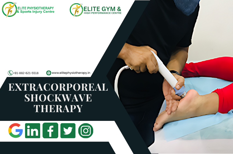
What is Extracorporeal Shockwave Therapy?

ESWT is a non-invasive procedure that uses acoustic waves to promote tissue healing, lessen discomfort, and increase range of motion. Originally developed to dissolve kidney stones, this method has been adapted to treat various sports and orthopedic conditions. Deeply penetrating the afflicted area, the high-energy sound waves encourage tissue regeneration, collagen synthesis, and blood circulation.

Conditions Treated with Shockwave Therapy

At Elite Physiotherapy and Sports Injury Centre, we use ESWT to treat a wide range of conditions, including:

  1. Plantar Fasciitis: Chronic heel discomfort brought on by plantar fascia inflammation is known as plantar fasciitis.
  2. Tennis and Golfer’s Elbow: Tendinopathies brought on by repeated strain on the elbow tendons are known as tennis and golfer’s elbow.
  3. Shoulder Calcific Tendonitis: Pain and stiffness are caused by calcium buildup in the shoulder tendons.
  4. Achilles Tendinopathy: Achilles tendinopathy causes pain and inflammation in the Achilles tendon.
  5. Patellar Tendinopathy: Patellar tendinopathy, commonly known as jumper’s knee, is a common condition in sports.
  6. Myofascial trigger pointsExcruciating muscular knots that limit range of motion.
  7. Shin Splints: Overuse or stress-related pain along the shin bone.

Shockwave therapy effectively treats many other sports and musculoskeletal injuries beyond the conditions listed above..

How ESWT Works

The therapy uses a specialized device to deliver focused or radial shockwaves to the affected area. These shockwaves accelerate the body’s natural healing processes by:

  • Increasing blood flow to the area.
  • Improving the mechanics of cellular repair.
  • Breaking down calcified deposits.
  • Desensitizing nerve endings to lessen pain.

The treatment requires no anesthesia or recovery time and takes place in brief sessions, typically lasting 15 to 20 minutes. After three to five sessions, the majority of patients report notable improvements.

Why Choose Elite Physiotherapy and Sports Injury Centre for ESWT?

To achieve the best outcomes, we at Elite Physiotherapy and Sports Injury Centre mix state-of-the-art equipment with individualized treatment. What makes us unique is this:

  1. Expert Team: Our physiotherapists stay updated on the latest ESWT procedures to deliver safe and efficient therapy.
  2. Holistic Approach: To address the underlying source of pain, we combine ESWT with other evidence-based treatments such as manual therapy, exercise rehabilitation, and recovery sessions like ice baths, hydrotherapy, etc.
  3. Patient-Centered Care: We customize each treatment program to match the patient’s needs, objectives, and lifestyle.
  4. Modern Equipment: Our clinic has state-of-the-art ESWT equipment, which guarantees accurate and effective care.

Benefits of ESWT

There are several advantages for patients receiving ESWT at Elite Physiotherapy and Sports Injury Centre, such as:

  • Pain relief: A notable decrease in persistent pain that doesn’t require medicine.
  • Faster Recovery: Injured tissues heal more quickly.
  • Better Mobility: Restoring function and movement in afflicted areas.
  • Non-Invasive: A risk-free, minimally invasive surgical substitute.

Your Journey to Pain-Free Living Starts Here

Extracorporeal shockwave therapy may provide the answer you need for chronic pain or a sports injury. Our goal at Elite Physiotherapy and Sports Injury Centre is to assist you in achieving both peak performance and long-lasting relief.

Contact us today to schedule a consultation and take the first step towards a healthier, pain-free life.

High-Intensity Class 4 Laser Therapy

At Elite Physiotherapy & Sports Injury Centre, we’re dedicated to offering our patients state-of-the-art care that will accelerate their recovery, improve their performance, and reduce their periods of inactivity. High-intensity Class 4 Laser Therapy (HILT) is one of our most sophisticated and successful techniques. This ground-breaking technology is revolutionizing the treatment of long-term conditions and sports injuries, providing athletes and individuals with unmatched outcomes.

What is High-Intensity Class 4 Laser Therapy?

High-Intensity Class 4 Laser Therapy is a non-invasive procedure that stimulates cellular repair and lessens pain by penetrating deeply into the tissues with concentrated light energy. The Class 4 laser has a higher power output than lower-class lasers, which allows it to more efficiently penetrate deeper tissue layers. This makes it the perfect option for managing chronic pain, sports injuries, and the healing process following surgery.

The Science Behind Class 4 Laser Therapy

Class 4 laser therapy’s therapeutic effects stem from a process known as photobiomodulation. When the laser light interacts with cells, it:

  1. Reduces Inflammation: By lowering the synthesis of pro-inflammatory mediators, the laser light helps to lessen swelling and encourage quicker healing.
  2. Boosts Cellular Metabolism: When cells produce more ATP (adenosine triphosphate), they are better equipped to heal damaged tissues.
  3. Enhances Blood Flow: Better microcirculation expedites the healing process by bringing nutrients and oxygen to the injured area.
  4. Enhances Cellular Metabolism: Increased collagen synthesis is necessary for tissue repair because it fortifies and repairs damaged tissues.

Benefits of High-Intensity Class 4 Laser Therapy at Elite Physiotherapy

  1. Fast Pain Relief: By focusing on pain receptors and lowering inflammation, the treatment offers both short-term and long-term pain relief.
  2. Faster Recovery: When compared to conventional techniques, HILT dramatically shortens the recovery period for sprains, muscle tears, and tendinitis.
  3. Drug-free and non-intrusive: This therapy is perfect for sports and provides a safe substitute for drugs and intrusive procedures.
  4. Customizable Treatments: Every patient can receive the best outcomes possible by modifying the Class 4 laser to meet their specific demands.

Applications in Sports and Rehabilitation

High-Intensity Class 4 Laser Therapy is utilized successfully at Elite Physiotherapy and Sports Injury Centre for:

  • Acute and Chronic Injuries: ranging from sprains of the ligaments to overuse injuries like runner’s knee and tennis elbow.
  • Post-Surgical Recovery: Quickening the healing process following orthopedic procedures like ACL replacement.
  • Muscle and Joint Pain: Managing ailments such as frozen shoulder, bursitis, and osteoarthritis.
  • Performance Optimization: Performance optimization is the process of helping top athletes recover and perform at their best.

Why Choose Elite Physiotherapy and Sports Injury Centre?

Our experience and individualized approach make our center unique. We incorporate High-Intensity Class 4 Laser Therapy into comprehensive treatment plans that are customized to meet the specific needs of each client, making us a center for excellence in sports physiotherapy. Our cutting-edge facilities and highly qualified physiotherapists guarantee that you receive top-notch care in a comforting setting.

A Success Story: Transforming Lives Through Laser Therapy

After only a few sessions of Class 4 Laser Therapy, one of our top athletes who had been recovering from a hamstring strain saw impressive results. The athlete returned to training weeks ahead of schedule after experiencing a significant reduction in pain and a return to mobility. This success story is only one of many that demonstrate how successful this cutting-edge therapy is.

Conclusion

High-Intensity Class 4 Laser Therapy is a revolutionary approach to managing and recovering from sports injuries. We at Elite Physiotherapy and Sports Injury Centre are pleased to provide our customers with this cutting-edge technology so they can live pain-free, perform better, and recover more quickly. Get in touch with us right now to discover the life-changing potential of Class 4 Laser Therapy if you’re recovering from an injury or want to speed up your healing.

Make an Appointment Now!
Learn how High-Intensity Class 4 Laser Therapy can improve your performance and recovery. Get in touch with Elite Physiotherapy and Sports Injury Centre to begin your journey to a more active, healthy lifestyle.

Baker’s Cyst and Its Comprehensive Management

Baker’s cyst, sometimes called a popliteal cyst, is a swelling behind the knee filled with fluid. It happens when synovial fluid builds up in the popliteal bursa, a little sac that lessens friction between the tissues around the knee joint. Although Baker’s cyst is usually not fatal, it can be extremely uncomfortable and restrict physical activity, particularly in athletes and athletic people.

Our specialty at Elite Physiotherapy and Sports Injury Centre is diagnosing and treating Baker’s cysts using advanced modalities and Evidence-based physiotherapy procedures to guarantee the best possible outcome.

Causes of Baker’s Cyst

Usually, an underlying knee condition leads to Baker’s cyst forming. Typical reasons include:

  • Osteoarthritis in the knee
  • Rheumatoid arthritis.
  • Meniscal tears
  • Damage to cartilage
  • Overuse injuries or trauma

For instance, a meniscal tear sustained by an elite athlete who jumps a lot could result in increased production of synovial fluid and the development of a Baker’s cyst.

Mechanism of Injury

When the knee joint becomes inflamed or irritated, too much synovial fluid is produced, which leads to the formation of the cyst. Through a one-way valve, this fluid escapes into the popliteal bursa. The bursa swells and develops into a palpable cyst behind the knee over time.

Signs, Symptoms, and Clinical Features of Baker’s Cyst

The following are Key symptoms of a Baker’s cyst:

  • Tightness and swelling behind the knee
  • Pain that gets worse when you flex your knees or stand for a long time
  • Knee with limited range of motion (ROM)
  • A bulge in the popliteal region that is felt or apparent
  • Discomfort or stiffness during exercising

In extreme situations, the cyst may burst, allowing fluid to seep into the calf and producing symptoms similar to deep vein thrombosis (DVT), including redness, swelling, and warmth.

Diagnostic Methods at Elite Physiotherapy

At Elite Physiotherapy and Sports Injury Centre, we employ a combination of physical and functional assessments to diagnose Baker’s cysts effectively.

Physical Assessment:

  • Check for obvious edema in the popliteal area.
  • palpation to determine the cyst’s presence and size.

Special Physiotherapy Tests for Baker’s Cyst:

  • The Bounce Home Test: Evaluates the knee joint’s integrity and detects any mechanical obstruction or effusion.
  • McMurray’s Test: Assesses meniscal damage that could be a factor in the development of cysts.
  • Apley’s Test: Assists in determining meniscal or ligamentous involvement.
  • Modified Thompson Test: In order to rule out other diseases like DVT, this test looks for fluid leaking into the calf.

Functional Assessment:

  • Examination of Gait patterns.
  • Assessment of joint stability and muscle strength.
  • To identify restrictions, use a range of motion testing.

Physiotherapy Management of Baker’s Cyst at Elite Physiotherapy

We treat Baker’s cysts using an individualized, evidence-based strategy that aims to minimize symptoms, enhance function, and stop recurrence. The cutting-edge treatments we provide are listed below:

1. Electrotherapy Modalities:

  • Shock Wave Therapy: Promotes tissue regeneration, lowers inflammation, and eases popliteal discomfort.
  • The Super Inductive System (SIS): Uses high-frequency electromagnetic stimulation to improve muscle activation and lessen knee stiffness.
  • High-Intensity Class IV Laser Therapy: Promotes the reabsorption of synovial fluid, reduces inflammation, and speeds up tissue recovery.

2. Hydrotherapy:

  • In a regulated setting, water-based workouts improve muscular strength and mobility while lowering joint stress.

3. Cryotherapy:

  • Localized cold therapy reduces pain and swelling, particularly after physical exercise.

4. Manual Therapy:

  • Soft tissue mobilization to ease constriction around the popliteal region.
  • Joint mobilization to increase knee range of motion.

5. Therapeutic Exercises for Baker’s Cyst:

  • Stretching exercises: Stretches for the hamstrings and calves help release tension in the area surrounding the knee joint.
  • Strengthening Exercises: To improve joint stability, concentrate on your quadriceps, hamstrings, and glutes.
  • Proprioception and Balance Training: Enhancing knee control and preventing more injuries are two benefits of proprioception and balance training.

6. Taping Techniques:

  • Kinesiology taping helps to ensure adequate alignment during exercises and lessens the pressure on the knee joint.

7. Progressive Rehabilitation Program:

  • Designed to meet the needs of each participant, this program aims to gradually restore full function and resume daily activities or sports.

Why Choose Elite Physiotherapy for Baker’s Cyst?

Our team at Elite Physiotherapy and Sports Injury Centre is dedicated to providing top-notch care through individualized treatment plans and state-of-the-art technologies. In addition to providing symptom alleviation, our all-encompassing strategy guarantees the long-term avoidance of Baker’s cysts and related knee disorders. With access to cutting-edge techniques including hydrotherapy, shock wave therapy, and super inductive systems, we offer unparalleled proficiency in treating musculoskeletal disorders and sports injuries.

Speak with one of our experts now to start along the path to pain-free mobility and peak performance. Your recovery is our top concern at Elite Physiotherapy!

IT Band Syndrome and Its Management

IT Band Syndrome, also known as Iliotibial Band Syndrome, is a common overuse injury that affects athletes, especially runners, bikers, and those who engage in repetitive lower-limb exercises. We at Elite Physiotherapy and Sports Injury Centre are experts at identifying and successfully treating this illness, enabling athletes to resume their best performance.

Causes of IT Band Syndrome

The iliotibial (IT) band is a thick band of connective tissue extending from the hip to the shinbone along the outside of the thigh is called the iliotibial (IT) band. When this band irritates or inflames as a result of excessive contact against the lateral femoral condyle, it can lead to IT Band Syndrome. Typical reasons include:

  • Overuse: Running, cycling, and rowing are examples of repetitive knee flexion and extension exercises.
  • Biomechanical imbalances: Include differences in leg length, weak hip abductors, or poor foot arch mechanics.
  • Inappropriate training methods: Include abrupt increases in mileage, effort, or footwear.
  • Tight IT Band or Surrounding Muscles: The glutes, quadriceps, or hamstrings are less flexible when the IT band or surrounding muscles are tight.

Mechanism of Injury

IT Band Syndrome can result from both on-field and off-field activity. For example, because of insufficient hip stability, a sprinter pushing off quickly during a race may overload their IT band, resulting in microtrauma and ultimately inflammation. Similarly, the IT band may rub too much against the femur if you cycle repeatedly while sitting at an incorrect height.

Signs, Symptoms, and Clinical Features

People with IT Band Syndrome frequently exhibit the following symptoms and indicators:

  • Running or cycling exacerbates lateral knee pain, which is usually acute or burning.
  • Sensitivity along the IT band, especially in the area next to the knee.
  • Localized inflammation or swelling on the knee’s lateral side.
  • Discomfort when sitting for extended periods of time or when going downstairs.
  • When moving, there is an audible snapping or popping sound on the outside of the knee.

Diagnostic Methods at Elite Physiotherapy

To identify the underlying cause of IT Band Syndrome, our diagnostic method combines specific physiotherapy tests with thorough physical and functional evaluations:

1. Physical Assessment:

  • Examination of posture and gait.
  • Palpation along the IT band to detect discomfort.
  • Assessing the hip, knee, and ankle joints’ range of motion and flexibility.

2. Functional Assessment:

  • Evaluation of the mechanics of cycling or running.
  • Hip abductors, glutes, and quadriceps strength tests.

3. Special Physiotherapy Tests:

  • Ober’s Test: Assesses the tightness of the IT band.
  • Noble Compression Test: When knee flexion and extension are performed, the Noble Compression Test detects discomfort or crepitus at the lateral femoral condyle.
  • Modified Thomas Test: The modified Thomas test evaluates hip flexor and IT band flexibility.

Physiotherapy Management at Elite Physiotherapy

We at Elite Physiotherapy and Sports Injury Centre provide a thorough physiotherapy technique that is customized to meet the needs of every patient. In addition to treating underlying biomechanical abnormalities and symptom relief, we also aim to avoid recurrence. This is how we go about it:

1. Pain Management:

  • Cryotherapy: Applying ice to alleviate pain and inflammation is known as cryotherapy.
  • Electrotherapy: Techniques such as TENS or ultrasound that aid in pain management and recovery.

2. Manual Therapy:

  • Soft Tissue Mobilization: Deep tissue massage to relieve tensor fasciae latae (TFL), glutes, and IT band tension.
  • Myofascial Release: Targeting trigger points along the IT band and surrounding muscles is known as myofascial release.
  • Joint Mobilization: Enhancing the ankle, knee, and hip joints’ alignment and range of motion is known as joint mobilization.

3. Stretching Exercises:

  • Stretches that increase the flexibility of the quadriceps, hamstrings, glutes, and IT band can be both dynamic and static.
  • Examples include: The standing hip flexor stretch, pigeon position, and the cross-body IT band stretch.

4. Strengthening Exercises:

Targeted workouts to improve stability and address muscular imbalances:

  • Clamshells: To strengthen the gluteus medius, use clamshells.
  • Lateral Band Walks: Strengthens the hip abductors with lateral band walks.
  • Single-Leg Deadlifts: Improves synergy between the glutes and hamstrings.
  • Step-ups: Increase the general strength and stability of the lower limbs.

5. Biomechanical Correction:

  • Postural corrections and gait retraining are used to maximize movement patterns.
  • Suggestions for appropriate footwear or, if required, orthotics.

6. Return-to-Sport Training:

  • Utilizing sport-specific drills to gradually resume running, cycling, or other sports.
  • Exercises involving plyometry and agility can improve performance while lowering the risk of injury.

Example: IT Band Syndrome in a Cyclist

Think of a professional cyclist who gets lateral knee pain when training hard. We would evaluate their pedaling mechanics and bike fit at Elite Physiotherapy. In addition to physiotherapy treatments, we would address concerns such as incorrect saddle height or cleat position. The cyclist can resume training pain-free and more effectively with the use of focused manual treatment, stretching, and strengthening exercises.

Why Choose Elite Physiotherapy?

At Elite Physiotherapy and Sports Injury Centre, we combine clinical expertise with state-of-the-art facilities to deliver personalized care. Whether you’re a professional athlete or a recreational enthusiast, our evidence-based approach ensures effective recovery and optimal performance. Visit us to experience the best in sports injury management!

PCL Injuries and Advanced Physiotherapy Management

Limiting the tibia’s excessive posterior translation with respect to the femur makes the posterior cruciate ligament (PCL) essential for knee stability. Despite being less frequently injured than the anterior cruciate ligament (ACL), PCL injuries can majorly impact knee function, particularly in sports and physically active people. To ensure the best possible rehabilitation and return to peak performance, we at Elite Physiotherapy & Sports Injury Centre specialize in evidence-based management of PCL injuries.

Causes and Mechanisms of PCL Injury

High-impact trauma or abrupt, powerful movements are common causes of PCL injuries. Typical reasons include:

  • Direct Trauma: A direct impact, such as striking the dashboard in an automobile accident, forces the anterior tibia posteriorly.
  • Sports Injuries: abrupt knee hyperextension or hyperflexion, which is typical in sports like basketball, football, and wrestling.

Example: Sports-Related Mechanism of Injury

A professional football player may land on a flexed knee while simultaneously feeling an opposing force on the shin when diving to rescue a ball. The PCL is overstressed by this hyperflexion and posterior tibial translation, which can result in a partial or total tear.

Signs, Symptoms, and Clinical Features

Individuals with a PCL injury may present with:

  • Mild to moderate stiffness and swelling.
  • Weight-bearing difficulties, particularly when engaging in knee-flexion activities.
  • A sensation of the knee “giving way” or becoming unstable.
  • Knee discomfort in the back, especially when squatting or going downstairs.
  • Loss of full range of motion.

Diagnostic Methods: Physical and Functional Assessment

At Elite Physiotherapy & Sports Injury Centre, we diagnose PCL issues using a thorough and methodical process. Our functional and physical evaluation consists of:

Patient History: A detailed account of the symptoms and mechanism of the injury.
Physical examination: Keeping an eye out for any indications of deformity, edema, or bruises.

Special Physiotherapy Tests:

  • Posterior Drawer Test:  measures the tibia’s posterior translation to evaluate the PCL’s integrity.
  • Sag Sign: When the knee is flexed to 90 degrees, it shows a posterior sagging of the tibia.
  • Quadriceps Active Test: When using the quadriceps, the Quadriceps Active Test measures the posterior tibial displacement.

Our specialists can determine the extent of the injury by combining these tests with functional evaluations.

Physiotherapy Management for PCL Injuries

Our approach to treatment at Elite Physiotherapy & Sports Injury Centre is centered on individualized, non-invasive rehabilitation that is catered to each patient’s need. Our advanced management of physiotherapy consists of:

1. Pain and Swelling Control

  • Compression and the use of ice packs to lessen inflammation.
  • Application of electrical techniques for tissue repair and pain management, such as ultrasonography or interferential treatment (IFT).

2. Joint Mobility and Range of Motion (ROM)

  • Activities with controlled passive and active assistance to regain joint mobility without putting undue strain on the ligament.
  • Utilizing joint mobilizations and other manual therapy procedures to restore mobility.

3. Strengthening and Muscle Activation

  • To improve knee stability and resist posterior tibial translation, strengthen your quadriceps.
  • Exercise your hamstrings carefully to prevent posterior strain on the PCL.
  • To enhance general biomechanics, use progressive resistance training for the hip and core muscles.

4. Neuromuscular Training

  • To regain dynamic stability, perform proprioceptive exercises with balancing boards, BOSU balls, or single-leg stance exercises.
  • Functional training adapted to the patient’s daily routine or sport.

5. Functional and Sport-Specific Rehabilitation

  • For athletes getting ready to resume play, plyometric drills, agility training, and sport-specific activities are recommended.
  • Introduction of pivoting and high-impact activities gradually under the guidance of experts.

5. Advanced Modalities

  • In chronic situations, dry needling or extracorporeal shockwave therapy (ESWT) are used to promote recovery.
  • Blood Flow Restriction (BFR) therapy reduces joint strain while providing effective strengthening.

Why Choose Elite Physiotherapy & Sports Injury Centre?

We take great satisfaction in offering state-of-the-art treatment for PCL injuries at Elite Physiotherapy. Modern technology, manual therapy, and a patient-centered rehabilitation strategy are all incorporated into our approach. Our goals are to improve overall performance and stop future incidents in addition to treating the injury.

By choosing Elite Physiotherapy & Sports Injury Centre, you gain access to:

  • Expert Assessment: Using specific physiotherapy tests, extensive diagnostic capabilities are available.
  • Personalized Care Plans: Interventions specifically designed to meet your rehabilitation objectives and activity level target your specific needs.
  • Advanced Techniques: The most recent evidence-based methods for the best results.

Our dedication to quality guarantees the best possible recuperation for athletes and people of all activity levels. For additional information on managing injuries, check out the blog area of our website or contact one of our professionals for a consultation right now. Allow us to assist you in returning to your passions with greater strength than before!

Patellofemoral Ligament Injuries and Their Management

Sports and other high-impact activities frequently result in patellofemoral ligament injuries, especially those involving the Medial Patellofemoral Ligament (MPFL) and Lateral Patellofemoral Ligament (LPFL). These ligaments are essential for keeping the patella (kneecap) stable when moving. Pain, decreased functionality, and patellar instability can result from injuries to these ligaments. At Elite Physiotherapy & Sports Injury Centre, we specialize in diagnosing and managing such injuries using cutting-edge technology and personalized care.

Understanding MPFL and LPFL

MPFL: The MPFL is the main stabilizer that keeps the patella from moving laterally. During patellar subluxations or dislocations, it frequently sustains injury.

LPFL: Although less frequently injured, the LPFL is essential for preserving medial patellar alignment.

Injuries, overuse, or anatomical predispositions such as muscle imbalances or malalignment can all cause injuries to these ligaments.

Causes and Mechanism of Injury

Causes

Trauma: Sudden twisting motions or direct impact to the knee.

Overuse: Constant tension brought on by exercises like jumping or running.

Anatomical factors: include a shallow trochlear groove, weak quadriceps, or patella malalignment.

Previous Dislocations: increased susceptibility following an initial injury.

Mechanism of Injury

MPFL Injuries: MPFL injuries are frequently brought on by lateral patella dislocation, which is frequently brought on by valgus force in conjunction with external knee rotation.

Example: A soccer player makes a sudden turn to avoid an opponent during a game. The patella dislocates laterally as a result of the planted leg undergoing an external rotation and the knee experiencing valgus stress. The MPFL, the main barrier preventing lateral patellar movement, is strained or torn by this force. Immediate pain, swelling, and an incapacity to play are experienced by the player.

This illustration demonstrates a typical on-field mechanism of injury including sudden direction changes, which are characteristic of sports like volleyball, basketball, and soccer.

LPFL Injuries: Injuries to the Lateral Patellofemoral Ligament (LPFL) are less common than those of the MPFL but can significantly affect knee stability and function. High-energy trauma or specific sports-related incidents that displace the patella medially often cause LPFL injuries.

Example: A valgus force is applied when a weightlifter’s knees fold inward during a heavy barbell squat because of poor form or muscle fatigue. The LPFL is strained or torn concurrently with an excessive medial pull on the patella brought on by hyperactive quadriceps or inadequate gluteal activation. The athlete has trouble bearing weight and immediately feels pain on the lateral side of the knee.

This illustration demonstrates that improper technique or muscular imbalances when lifting weights can cause LPFL injuries.

Signs and Symptoms

Pain: Located near the patella, frequently made worse by stair climbing and squatting.

Swelling: Acute swelling brought on by harm to soft tissues.

Instability: The sensation that the knee is “giving way,” particularly when moving laterally.

Reduced Range of Motion (ROM): Inability to bend or extend.

Tenderness: Palpating the MPFL or LPFL regions reveals noticeable tenderness.

Patellar maltracking: Patellar maltracking refers to a visual or tactile deviation during movement.

Diagnosis at Elite Physiotherapy

Clinical and Functional Assessment

Our diagnostic methodology incorporates specialized physiotherapy tests .

Observation: Look for quadriceps atrophy, edema, and patellar malalignment.

Palpation: Determine whether an area is LPFL or MPFL tender.

Special Tests

  • Patellar Apprehension Test: A positive patellar apprehension test is indicated when a patient exhibits apprehension during lateral patellar translation, suggesting an MPFL injury.
  • Moving Patellar Tracking Test: The Moving Patellar Tracking Test detects abnormalities in the patellar glide.
  • Medial Patellofemoral Ligament Stress Test: Our Team evaluates the MPFL’s integrity using the medial patellofemoral ligament stress test.

Physiotherapy Management for Patellofemoral Ligament Injuries

To handle such conditions, Elite Physiotherapy & Sports Injury Centre uses a thorough, individualized approach that incorporates cutting-edge methods and the latest equipment to guarantee the best possible recovery. Here’s a detailed look into the physiotherapy protocol:

1. Initial Assessment and Goal Setting

Detailed Evaluation: Determine the severity of the injury, patellar tracking, and any underlying causes, such as biomechanical problems or muscle imbalances.

Goal Setting: Setting goals that are specific to the requirements of athletes or active people should center on reducing pain, regaining stability, and avoiding recurrence.

2. Pain Management and Early Rehabilitation

Modalities: Methods for reducing pain and inflammation, such as laser therapy, shockwave therapy, and cryotherapy.

Immobilization and Protection: Our Expert Physiotherapist may advise temporary bracing to stabilize the knee.

3. Restoring Mobility and Strength

Range-of-motion (ROM) exercises, including passive and active: Start with controlled motions to protect the injured ligament and avoid stiffness.

  • Muscle Strengthening:
  • Quadriceps: To improve patellar tracking, concentrate on the Vastus Medialis Oblique (VMO).
  • Gluteal muscles: improve stability in the hips.
  • Core Strengthening: Strengthening your core will increase your overall functional stability.

4. Proprioception and Neuromuscular Training

Use of balance boards, Proprioceptive exercises, and cutting-edge equipment such as Neuromuscular Electrical Stimulation (NMES) are used to retrain knee stability and coordination.

5. Advanced Functional Training

gradual return to sports-specific motions with the aid of resistance training, hydrotherapy, and Kinesio Taping.

Sport-Specific Drills: A focus on agility training, plyometric activities, and return-to-sport procedures for top athletes.

6. Preventive Strategies and Education

Correct predisposing variables, including incorrect footwear or training mistakes.

self-management skills along with stretching and warm-up activities.

Why Choose Elite Physiotherapy & Sports Injury Centre?

To ensure a quicker recovery and the best possible outcomes, Elite Physiotherapy blends evidence-based procedures with state-of-the-art technologies including Extracorporeal Shockwave Therapy (ECSWT), Cryo-Air Therapy, and High-Intensity Class IV Laser Therapy. The center’s comprehensive rehabilitation strategy helps patients recover from injuries and perform at their best.​

For more insights or to book a consultation, visit the Elite Physiotherapy & Sports Injury Centre.

Patellofemoral Instability: And its Physiotherapy Management

Incorrect alignment or frequent patella (kneecap) dislocation concerning the femoral groove (trochlea) is a complex problem known as patellofemoral instability. If left untreated, this condition may result in chronic joint degeneration, pain, and functional restrictions. Whether they are athletes, active people, or anybody else coping with patellofemoral issues, our goal at Elite Physiotherapy and Sports Injury Centre is to offer the best care possible.

Anatomy of the Patellofemoral Joint

One of the knee’s essential parts is the patellofemoral joint. When walking, jogging, or kneeling, the patella moves smoothly inside the femur’s trochlear groove. Stability is preserved by:

  • Bony structures: The patella’s form and the depth of the trochlear groove.
  • Soft tissue restraints: The patellar tendon, quadriceps muscles, lateral retinaculum, and medial patellofemoral ligament (MPFL) are examples of soft tissue constraints.

When these components become disorganized or imbalanced, instability may result, which might cause the patella to move laterally or dislocate entirely.

Causes of Patellofemoral Instability

Traumatic Events: Patellar dislocation is frequently caused by direct strikes or twisting injuries.

Anatomical Predispositions:

  • trochlear groove that is shallow.
  • High-riding patella, or patella alta.
  • The Q-angle, or the angle formed by the patellar tendon and quadriceps.

Muscle Imbalances:

  • Weakness or delayed vastus medialis obliquus (VMO) activation.
  • tight lateral structures, such as the lateral retinaculum or iliotibial band.

Ligamentous Laxity: People who have conditions like Ehlers-Danlos syndrome may be more prone to instability in the future.

Recurrent Dislocations: The patella is more vulnerable to instability in the future if it has been dislocated.

Symptoms of Patellofemoral Instability

Patients frequently arrive with:

  1. Knee Pain: Knee pain is mostly anterior and is worse when you squat or climb stairs.
  2. Instability:  An unstable knee feels like it’s “giving way.”
  3. Swelling: Particularly following a first displacement.
  4. Reduced Range of Motion: As a result of mechanical impediments or pain.
  5. Apprehension Sign: Fear or discomfort when the patella is moved laterally is an indication of apprehension.

Diagnosis

At Elite Physiotherapy and Sports Injury Centre, we evaluate patellofemoral instability using a variety of cutting-edge diagnostic techniques:

Clinical Assessment:

  • Palpation to find effusion or discomfort.
  • Special tests such as the J-sign observation, apprehension test, and patellar glide test.

Apart from our assessment, patellar instability can be diagnosed also by:

Imaging:

  • X-rays: To evaluate trochlear morphology and bone alignment.
  • MRI: For assessing soft tissues, such as cartilage degeneration and MPFL integrity.
  • CT scans: To evaluate rotational alignment and measure patellar tilt.

Treatment Options

Non-Surgical Management

The first line of treatment is frequently non-surgical, particularly for people who have had only one dislocation or who have modest instability.

Physiotherapy:

The mainstay of treatment for patellofemoral instability is physiotherapy, which aims to enhance patellar tracking, stabilize the joint, and allow for a full return to function. We use a thorough and sophisticated approach at Elite Physiotherapy & Sports Injury Centre, combining manual therapy, therapeutic exercises, and state-of-the-art modalities.

Key Components of Physiotherapy Management
1. Management of Pain and Inflammation
  • Cryotherapy: Using ice packs to minimize swelling and lessen pain.
  • High-Intensity Laser Therapy (HILT): By promoting cellular regeneration, high-intensity laser therapy (HILT) speeds up tissue healing, lowers inflammation, and offers profound pain relief.
  • Shock Wave Therapy: Shock wave therapy is useful for improving the repair of the soft tissues surrounding the patellofemoral joint and reducing chronic pain.
2. Soft Tissue and Joint Mobilization

Manual Therapy:

  • Soft tissue mobilization: For tight lateral structures like the lateral retinaculum and iliotibial band, manual therapy involves mobilizing soft tissues.
  • patellar mobilizations: To enhance trochlear groove alignment and gliding.

Cupping therapy: improves circulation and eases knee-related muscle strain.

3. Neuromuscular Activation and Muscle Strengthening
  • To improve patellar tracking, the quadriceps should be strengthened, especially the vastus medialis obliquus (VMO).
  • To improve total knee stability, progressive resistance training focuses on the calf, gluteal, and hamstring muscles.
  • deep neuromuscular activation, which enhances muscle recruitment and decreases spasticity, through the use of advanced equipment like the Super Inductive System (SIS).
4. Stretching and Flexibility Training
  • To ease tightness and encourage balanced patellar movement, perform stretches for the lateral structures, quadriceps, hip flexors, and iliotibial band.
  • methods for increasing flexibility, such as Proprioceptive Neuromuscular Facilitation (PNF) stretching.
5. Proprioception and Balance Training
  • To improve neuromuscular coordination and joint proprioception, use stability balls, wobble boards, and balancing pads.
  • Functional exercises and sport-specific drills to enhance dynamic stability during intricate motions.
6. Advanced Physiotherapy Modalities
  • Capacitive-Resistive Energy Transfer (C.R.E.T.) therapy: A non-invasive method that enhances tissue circulation and oxygenation, decreases inflammation, and speeds up recovery.
  • Shock Wave Therapy: Encourages soft tissue healing, increases blood flow, and dissolves calcifications.
  • The Super Inductive System (SIS): Uses high-frequency electromagnetic stimulation to help with pain alleviation and muscular strengthening.
7. Functional Rehabilitation
  • Including lunges, step-ups, and squats while paying close attention to proper patellar monitoring.
  • A gradual return to dynamic exercises, such as jumping and sideways motions, to maintain control and alignment when doing sports-specific duties.
8. Patellar Stabilization Techniques
  • Taping: To offer support and adjust patellar alignment, use McConnell or kinesiology taping techniques.
  • Bracing: Personalized patellar braces to reduce excessive movement and boost self-assurance while engaging in exercises.
9. Patient Education
  • Education on good posture, movement patterns, and biomechanics to reduce excessive knee strain.
  • Recurrence prevention guidelines for safe sports and activity participation.

Surgical Management

Recurrent instability, severe anatomical anomalies, or the failure of conservative therapy may all require surgery. Typical procedures consist of:

  • MPFL Reconstruction: Rebuilding the ligament to stop the patella from moving laterally is known as MPFL reconstruction.
  • Trochleoplasty: When a patient’s trochlea is flat, the trochlear groove is deepened.
  • Tibial Tubercle Transfer: Patellar tendon realignment for proper tracking.
  • Lateral Release: Reducing patellar tilt by cutting tight lateral structures.

When surgery is necessary, Elite Physiotherapy and Sports Injury Centre will work post-operative management to provide a smooth transition from surgery to recovery.

At Elite Physiotherapy and Sports Injury Centre, we incorporate these therapies into a comprehensive treatment program that is tailored to each patient’s specific requirements. Our blend of state-of-the-art modalities and evidence-based practices guarantees the best possible recovery and long-term avoidance of patellofemoral instability.

Jumper’s Knee: Understanding and Managing Patellar Tendinopathy

A frequent overuse condition that affects the patellar tendon, which joins the kneecap (patella) to the shinbone (tibia), is known clinically as jumper’s knee or patellar tendinopathy. Athletes who play high-impact sports like basketball, volleyball, and soccer that entail repetitive jumping are more likely to have this medical condition. We at Elite Physiotherapy and Sports Injury Centre are experts at treating jumper’s knee, integrating cutting-edge therapeutic approaches with individualized rehabilitation plans to promote speedy recovery and avoid recurrence.

Anatomy and Mechanism of Injury

Activities like jumping, running, and landing are made possible by the patellar tendon, which transfers force from the quadriceps muscle to the lower leg. When a tendon experiences repeated stress and pressure, it can develop microtears, inflammation, and eventually degeneration if treatment is delayed. This condition is known as jumper’s knee. This tension eventually results in knee pain, swelling, and decreased function.

Symptoms of Jumper’s Knee

Typical symptoms of jumper’s knee include:

  • pain near the bottom of the kneecap, particularly during exercising or just after.
  • Tenderness at the patellar tendon.
  • Swelling and localized warmth around the knee.
  • Stiffness or a “tight” sensation, especially after rest.
  • Reduced Performance in jumping and squatting due to pain.

If treatment is not received, the symptoms will eventually impair an athlete’s ability as they frequently get worse with sustained exertion.

Causes and Risk Factors

A number of causes can contribute to jumper’s knee, including:

  • Overuse and High Training Volume: Repeated jumping and landing overstress the patellar tendon.
  • Muscle Imbalances: Weakness in the hip, hamstring, or quadriceps muscles can increase tendon stress.
  • Poor Technique: Improproper landing or jumping movements can increase impact pressures on the knee.
  • Poor Surface and Footwear: Hard surfaces or worn-out shoes are unable to absorb trauma, which can lead to tendon overload.
  • Inadequate Recovery: Tendons take longer to repair after strenuous exercise, which raises the risk of injury.

Diagnosis at Elite Physiotherapy and Sports Injury Centre

At Elite Physiotherapy and Sports Injury Centre, we diagnose jumper’s knee using a thorough process. If necessary, healthcare providers perform diagnostic imaging, such as an MRI or ultrasound, in addition to conducting a comprehensive physical examination that includes evaluating knee function and palpation. This aids in diagnosing the condition and determining the degree of tendon involvement, which helps us create a customized rehabilitation program.

Physiotherapy Management of Jumper’s Knee

We can effectively manage jumper’s knee through various methods, focusing on activity modification, tendon repair, pain management, and strength rebuilding. Elite Physiotherapy and Sports Injury Centre uses the following evidence-based treatments:

  1. Management of Pain and Inflammation

Cryotherapy, also known as cold treatment, helps to lessen pain and inflammation.

Electrotherapy Modalities: Methods like TENS (Transcutaneous Electrical Nerve Stimulation) and ultrasound therapy can provide pain relief and promote healing.

Activity Modification: To give the tendon time to heal, it is crucial to cut back on high-impact exercises like leaping, squatting, and running.

  1. Tendon Rehabilitation with Eccentric Exercises

Eccentric Strengthening Protocols: To gradually stress the muscles and promote tendon remodeling, people use eccentric activities like slow lunges and decline squats.

Isometric Exercises: In the early stages of rehabilitation, we use isometric exercises to build strength and relieve discomfort without placing excessive strain on the tendons.

  1. Quadriceps and Hip Strengthening

Quadriceps Strengthening: By strengthening the quadriceps specifically, a program can lessen the strain on the patellar tendon.

Exercises for Hip Stabilization: Maintaining hip stability and strength is essential for managing knee mechanics. The program includes exercises like clamshells, hip bridges, and hip abductor strengthening.

  1. Corrective Exercises and Movement Re-education

Biomechanical Analysis: By evaluating the athlete’s jumping and landing motions, we can identify defects that increase stress on the knee. We use plyometric exercises and movement retraining to address these habits.

Soft Tissue Techniques and Myofascial Release: We use deep tissue release and foam rolling techniques to relieve tension in the knee muscles, ensuring improved range of motion and flexibility.

  1. Gradual Return to Activity

Activity-Specific Drills: To get the athlete ready to resume full activity, we gradually introduce sport-specific drills as the tendon strengthens. This approach allows people to regain their performance and confidence without risking re-injury.

Education on Load Management: To reduce the chance of recurrence, our staff offers advice on how to manage training loads.

Advanced Treatment Options Available at Elite Physiotherapy and Sports Injury Centre

We may recommend other therapies for athletes with chronic or persistent conditions:

Shockwave Therapy: By applying acoustic waves to the injured region, this technique increases blood flow and aids in tendon recovery.

CRET Therapy: CRET therapy promotes blood flow and cellular healing by producing deep heat in the tendon. By promoting tissue regeneration, it lessens discomfort and hastens the healing process for jumper’s knee.

Class 4 Laser Therapy: By delivering concentrated laser energy deep into tissues, class 4 laser treatment lowers inflammation and promotes tendon healing. Additionally, it reduces pain, making it easier for athletes with jumper’s knee to advance through their recovery.

Super Inductive System (SIS) Therapy: It stimulate muscles and encourage neuromuscular re-education. It is an excellent, non-invasive therapy option for jumper’s knee since it stimulates blood flow, relieves pain, and improves knee stability.

Bracing and Taping Techniques: Patellar straps and taping techniques help relieve the tendon’s tension while you’re exercising.

Prevention of Jumper’s Knee

Athletes who want to continue performing at a high level must avoid jumper’s knee. Our emphasis at Elite Physiotherapy and Sports Injury Centre is on thorough conditioning regimens that consist of:

Frequent Strength and Flexibility Training: Supporting knee mechanics requires balanced training of the hips, hamstrings, and quadriceps.

Training in Biomechanics and Technique: Reducing impact forces requires proper landing and jumping skills. Specifically, mastering these techniques can help minimize stress on the joints and prevent injuries.

Rehabilitation Strategies: To avoid overusing tendons, we recommend getting enough sleep and incorporating rehabilitation techniques, such as foam rolling and stretching. In addition, these practices help promote recovery and reduce the risk of injury.

Why Choose Elite Physiotherapy and Sports Injury Centre?

Our strategy for treating jumper’s knee at Elite Physiotherapy and Sports Injury Centre is based on knowledge, individualized treatment, and evidence-based rehabilitation. We collaborate closely with each athlete to create a personalized recovery plan that takes into account their particular requirements and sporting objectives. Our staff ensures that each patient is informed about their condition, available treatments, and preventive measures to empower them on their path to full recovery and enhanced performance.

By choosing us, athletes may receive the best care possible and access a variety of cutting-edge treatment techniques, which will help them recover from injuries and return to their best performance.

Knee Arthritis

Millions of individuals worldwide suffer from knee arthritis, a degenerative joint disease that is more common among sports, older adults, and persons in high-impact jobs. For those with knee arthritis, our specialized therapeutic methods at Elite Physiotherapy and Sports Injury Centre focus on minimizing discomfort, regaining function, and enhancing quality of life. This article goes into great detail on knee arthritis, including its causes, symptoms, diagnosis, and the cutting-edge physiotherapy treatments our clinic offers.

What is Knee Arthritis?

The primary cause of knee arthritis is the degeneration of the knee joint’s cartilage, which results in pain, swelling, and restricted movement. Rheumatoid arthritis (RA) and osteoarthritis (OA) are the two most common types:

  • Osteoarthritis (OA): A “wear-and-tear” form of arthritis, osteoarthritis (OA) is frequently observed in athletes and older persons. It is caused by the slow deterioration of cartilage.
  • Rheumatoid Arthritis (RA): The autoimmune disease known as rheumatoid arthritis (RA) causes inflammation and potential joint abnormalities when the body’s immune system targets the joint lining.

Causes and Risk Factors

Numerous variables, including a mix of lifestyle, injury history, and genetics, can lead to knee arthritis. Important elements consist of:

Age:

  • As people age, especially beyond fifty, their risk of having knee arthritis increases.

Prior Injuries:

  • Knee injuries including meniscus tears, ligament damage, or fractures might raise the risk.

Weight:

  • Carrying too much weight strains the knee joints and speeds up cartilage deterioration.

Genetics:

  • People may be more susceptible to knee arthritis if they have a family history.

Occupational Factors:

  • Jobs involving heavy lifting or kneeling that cause repeated knee stress may increase the risk.

Symptoms of Knee Arthritis

The following symptoms are frequently seen in patients who arrive to Elite Physiotherapy and Sports Injury Centre:

Pain: Usually gets better with rest and gets worse with activities.

Swelling: Knee joint inflammation is frequent, particularly with extended usage.

Stiffness: It’s common to have less range of motion and flexibility, especially after extended periods of inactivity.

Deformity: Joint deformity may develop in more severe instances, impacting walking and alignment.

Grinding Sensation: When moving the knee, patients may feel as though it is cracking or grinding.

Seeking early assistance can help prevent long-term harm if you encounter any of these symptoms.

Diagnosis of Knee Arthritis

At Elite Physiotherapy and Sports Injury Centre, our team starts with a comprehensive clinical evaluation, which is frequently followed by imaging if required:

Clinical Examination: To determine the degree and functional limits, we evaluate alignment, muscular strength, and range of motion.

X-Rays: An X-ray can reveal the degree of bone alterations, joint space constriction, and cartilage loss.

MRI: To check for injury to the meniscus and ligaments, an MRI may be suggested for a thorough evaluation of soft tissues.

Physiotherapy Management at Elite Physiotherapy and Sports Injury Centre

One of the best non-invasive ways to treat knee arthritis is through physiotherapy. We provide customized programs at Elite Physiotherapy and Sports Injury Centre to promote joint function, mobility, and long-term pain reduction. The following therapies are incorporated into our approach:

1. Therapeutic Exercise

The foundation of managing arthritis is therapeutic exercise. To strengthen the muscles surrounding the knee, lessen joint stress, and promote general function, we employ specialized training regimens. Typical workouts consist of:

  • Quadriceps Strengthening: Strengthening the quadriceps with exercises like leg lifts and squats helps to stabilize the knee.
  • Hamstring and Glute Strengthening:  Such as bridges, can support the knees.
  • Range-of-Motion Exercises:  To preserve knee mobility, use mild stretching exercises.
  • Aerobic Exercises: Without putting undue strain on the knee joint, low-impact sports like swimming and cycling can help control weight and enhance cardiovascular fitness.

2. Manual Therapy

To increase blood flow, decrease stiffness, and move the knee joint, our licensed therapists use manual therapy:

  • Soft Tissue Mobilization: Lessens tenseness in the tendons and muscles around it.
  • Joint mobilizations: To increase the range of motion in the knee.
  • Myofascial Release: Pain relief and increased soft tissue flexibility are two benefits of myofascial release.

3. Electrotherapy

We use cutting-edge electrotherapy methods to reduce pain and manage inflammation, such as:

  • Ultrasound Therapy:  Promotes tissue repair by lowering pain and inflammation.
  • Transcutaneous Electrical Nerve Stimulation (TENS): By activating sensory nerves, transcutaneous electrical nerve stimulation (TENS) lessens the experience of pain.
  • Interferential Therapy (IFT): Low-frequency electrical currents are used in interferential therapy (IFT) to lessen inflammation and discomfort.
  • High-intensity class 4 laser: It helps to improve blood circulation to your knee which eventually helps in pain reduction and recovery.
  • CRET Therapy: It is a Highly advanced Device for Pain relief and faster recovery, it will help with knee arthritis and make your recovery faster.

4. Cryotherapy and Thermotherapy

Therapies focused on temperature can offer substantial relief:

  • Cryotherapy (Cold Therapy): Cryotherapy, often known as cold treatment, reduces swelling and inflammation, particularly after physical exertion.
  • Thermotherapy (Heat Therapy): Heat treatment, or thermotherapy, improves blood flow, eases muscular tension, and reduces stiffness.

Depending on each patient’s demands, our team frequently switches between cryotherapy and thermotherapy, using each in a specific manner to reduce symptoms.

5. Kinesiology Taping 

  • Kinesiology tape is one supportive approach that can relieve knee joint tension and increase stability.

6. Hydrotherapy (Water Therapy)

  • Exercises with water offer a low-impact, efficient way to strengthen and mobilize the knee without putting undue strain on it. For more severe forms of arthritis, this treatment is very helpful since it permits pain-free complete range of motion.

Lifestyle and Home Management Tips

Our goal at Elite Physiotherapy and Sports Injury Centre is to equip our patients with the knowledge they need to take care of their conditions daily. We provide advice on:

Weight management: Maintaining a healthy weight is essential for minimizing pressure on the knee joint.

Activity Modification: By offering guidance on how to modify everyday routines, our therapists assist patients in avoiding strenuous activities that worsen arthritis.

Pain management: Strategies include the use of walking aids, knee braces, and good posture.

Why Choose Elite Physiotherapy and Sports Injury Centre?

With our expertise in orthopedic and sports physiotherapy, Elite Physiotherapy and Sports Injury Centre is ready to provide specialized care for knee arthritis. You may rely on us for your recovery for the following reasons:

Expert Team: Our knowledgeable team of physiotherapists is current with the most recent findings and methods.

Customized Treatment Plans: We adjust each therapy program according to the patient’s objectives, activity level, and arthritis severity.

State-of-the-art Facilities: Our clinic has a variety of rehabilitation equipment, hydrotherapy pools, and advanced electrotherapy devices.

Patient-Centered Approach: From evaluation to discharge, we are dedicated to our patients’ recovery journey, ensuring they have a knowledgeable and supportive experience.

Conclusion

Although knee arthritis can be debilitating, it doesn’t have to define your life. Relief and increased functionality are achievable with the correct management strategy and a committed team at your side. Our goal at Elite Physiotherapy and Sports Injury Centre is to provide you with individualized, evidence-based care so you may live your best life.

Don’t put off getting assistance if you or someone you know has knee arthritis. To start your path to pain relief and increased mobility, get in touch with Elite Physiotherapy and Sports Injury Centre right now.

Chondromalacia Patella: Physiotherapy Management at Elite Physiotherapy and Sports Injury Centre

The common condition known as “chondromalacia patella,” or runner’s knee, damages the cartilage on the underside of the patella (kneecap). The primary cause of this condition is the weakening and degradation of the cartilage, which results in knee joint discomfort, inflammation, and dysfunction. Our specialty at Elite Physiotherapy and Sports Injury Centre treats these knee-related conditions with focused physiotherapy treatments meant to lessen pain, restore function, and stop recurrence. To properly treat chondromalacia patella, this article examines the cause, symptoms, diagnosis, and physiotherapy-focused treatment approaches.

1. Anatomy and Pathophysiology of Chondromalacia Patella

One of the most important parts of knee biomechanics is the patella or kneecap. It acts as a lever arm to boost the mechanical advantage of the quadriceps, particularly while squatting, sprinting, and jumping. Smooth movement along the femoral groove is made possible by the articular cartilage covering the bottom of the patella. Chondromalacia patella occurs when this cartilage weakens and degrades.

This deterioration frequently results from:

  • Overuse and Repetitive Stress: Athletes and anyone who engage in high-impact activities frequently experience overuse and repetitive stress.
  • Biomechanical Imbalances: Poor alignment, weak quadriceps, or hyperactive lateral thigh muscles are examples of biomechanical imbalances.
  • Injury or Trauma: Damage to the knee may result from a fall or a direct hit.
  • Aging: People may be more susceptible to cartilage degeneration as a result of normal wear and tear over time.

2. Symptoms and Clinical Presentation

Chondromalacia patella symptoms include:

  • Knee pain: particularly while jogging, bending, kneeling, or climbing stairs.
  • Tenderness and Swelling: Around the kneecap, which might be painful to the touch.
  • Grinding or Clicking Sensation: When the knee is bent or extended, there may be a grinding or clicking sensation.
  • Reduced Functionality: Instability and pain make it difficult to carry out daily tasks or participate in sports.

3. Diagnosis of Chondromalacia Patella

Our diagnostic process at Elite Physiotherapy and Sports Injury Centre combines a thorough physical examination, patient history, and, if required,  imaging methods.

Physical examination:

Evaluation of patellar tracking, muscular strength, knee range of motion, and any indications of pain.

The Patellar Compression Test:

This test measures the sensitivity of the cartilage beneath the kneecap and aids in reproducing pain.

Imaging (MRI):

By revealing the degree of cartilage destruction, MRI enables a targeted therapy strategy. X-rays can help to rule out other abnormalities of the bones.

4. Physiotherapy Management at Elite Physiotherapy and Sports Injury Centre

The mainstay of treatment for chondromalacia patella is physiotherapy. Our goal at Elite Physiotherapy and Sports Injury Centre is to provide each patient with tailored care that promotes the best possible knee health via strengthening, stretching, and functional rehabilitation. The following are the main components of our strategy:

A. Inflammation Control and Pain Management

Cryotherapy and Thermotherapy: Ice packs are first used in cryotherapy and thermotherapy to lessen pain and inflammation. Heat treatment can help to enhance blood flow and encourage tissue repair as the healing process advances.

Manual Therapy: To enhance patellar tracking and lessen pain, methods including soft tissue release and patellar mobilizations are used.

B. Strengthening the Quadriceps

Closed Kinetic Chain Exercises:  such as leg presses and squats, can strengthen the quadriceps while reducing the strain on the patella.

Isometric Quadriceps Contractions: In the early phases of rehabilitation, it is essential to maintain strength without putting undue strain on the joints. This can be achieved by isometric quadriceps contractions.

Straight Leg Raises and Short-Arc Quads: These exercises assist avoid overuse of the knee joint by engaging the quadriceps in a controlled way.

C. Correcting Biomechanical Imbalances

Strengthening the Hip Musculature: Patellar maltracking can result from improper knee alignment caused by weak hip abductors and rotators. To target these muscles, we concentrate on exercises like side-lying leg raises, hip bridges, and clamshells.

Stretching Tight Muscles: To increase flexibility and lessen aberrant patellar forces, foam roll and stretch the quadriceps, hamstrings, and iliotibial (IT) band.

D. Improving Patellar Tracking

Taping Techniques: Kinesiology and McConnell taping are used to stabilize the patella, allowing proper alignment during exercises and reducing pain.

Patellar Mobilization: Patellar mobilization refers to manual techniques that increase patellar mobility and prevent tightness, which can worsen chondromalacia symptoms.

E. Returning to Sport and Functional Training

Proprioceptive and Balancing Training: We improve joint stability and proprioception, which are essential for athletes and active people, by using Bosu balls, balancing boards, and single-leg exercises.

Gradual Reintroduction to Activity: Under supervision, patients gradually return to sport-specific exercises, ensuring a safe and effective recovery without re-injury.

5. Preventive Measures for Long-Term Management

Long-term healing depends on preventing recurrence. Elite Physiotherapy and Sports Injury Centre places a strong emphasis on the following:

  • Regular Strengthening and Conditioning: Maintaining knee stability requires regular hip and quadriceps strengthening.
  • Proper Footwear and Orthotics: To avoid further knee pain, those with flat feet or atypical foot mechanics may benefit from custom orthotics.
  • Education on Activity Modification: We advise patients on how to avoid putting too much strain on their knees, maintain good body mechanics, and set appropriate activity limitations.

6. Why Choose Elite Physiotherapy and Sports Injury Centre?

Our center is committed to providing comprehensive, science-backed treatment that addresses both the symptoms of chondromalacia patella and its underlying causes. Our staff adeptly uses cutting-edge physiotherapy techniques customized to meet each person’s unique health and lifestyle needs. In addition to pain management, we strive for long-term prevention and functional enhancement so that our patients may confidently resume their activities.

At Elite Physiotherapy and Sports Injury Centre, we support a patient-centered strategy that blends individualized care with professional knowledge. Contact our knowledgeable team for a consultation and start your path to pain-free movement if you or a loved one is experiencing knee discomfort or has been diagnosed with chondromalacia patella.