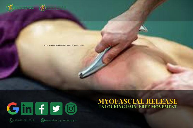
Cupping Therapy: The Secret to Faster Muscle Recovery

Introduction

An age-old healing technique, cupping therapy has become more relevant in physiotherapy nowadays. It improves circulation, eases tense muscles, and speeds up the healing process by using suction. For faster recovery and enhance athletic performance, we at Elite Physiotherapy and Sports Injury Centre combine evidence-based physiotherapy with cupping.

What Is Cupping Therapy?

In order to create suction, specialized cups are applied to the skin during cupping therapy. Soft tissues are raised by this negative pressure, which also encourages blood flow to the intended location. This method is used by physiotherapists to efficiently treat muscle limitations, discomfort, and stiffness.

Types of Cupping Techniques

  • Dry Cupping: This method involves using suction without making any cuts. It eases muscle tension and improves tissue mobility.
  • Wet Cupping: To remove stagnated blood, superficial skin pricks are performed prior to suction.
  • Dynamic or Sliding Cupping: To increase soft tissue elasticity and loosen fascia, cups are moved across lubricated skin.
  • Fire Cupping: The old-fashioned technique of “fire cupping” uses heat to generate suction inside the cups.

Dynamic and dry cupping are most frequently utilized at Elite Physiotherapy because of their efficacy and safety in sports rehabilitation. Also we Practice Wet cupping in a very professional and hygienic environment which is performed by our skilled therapists

Mechanism of Action

By producing negative pressure, cupping therapy raises underlying tissues and improves local blood flow. This increases the flow of oxygen and speeds up the elimination of waste products from metabolism. Additionally, it stimulates the parasympathetic nervous system, which aids in rest and recovery.

Benefits of Cupping Therapy in Physiotherapy

  1. Reduces Muscle Pain: Suction eases tense muscles and trigger points.
  2. Enhances Blood Flow: Improved circulation supports tissue repair.
  3. Encourages Fascia Release: By releasing fascial adhesions, this technique aids in the restoration of regular movement patterns.
  4. Reduces Inflammation: Facilitates the elimination of toxins and lymphatic drainage.
  5. Increases Flexibility: Increases range of motion and decreases stiffness.
  6. Accelerates Recovery: Perfect for athletes recovering from training or injuries.

At Elite Physiotherapy and Sports Injury Centre, physiotherapists combine cupping with advanced techniques such as the Super Inductive System (SIS), CRET Therapy, Shock Wave Therapy, and Class 4 Laser for faster results.

Clinical Applications of Cupping Therapy

Numerous musculoskeletal and sports injury issues can benefit from cupping therapy, such as:

  • Myofascial pain syndrome
  • Rotator cuff strain and shoulder impingement
  • SI joint dysfunction together with lower back discomfort
  • Tightness in the calf and hamstrings
  • IT band friction syndrome
  • Post-exercise muscle soreness
  • Chronic neck and upper back stiffness

At Elite Physiotherapy, physiotherapists customize every session after a thorough evaluation to ensure safe and effective treatment.

Safety and Precautions

When administered by qualified physiotherapists, cupping therapy is safe. Circular markings or mild redness are common and go away in a few days. Avoid cupping therapy in cases of pregnancy, open wounds, bleeding disorders, and skin infections.

Every session at Elite Physiotherapy adheres to stringent safety and hygiene regulations.

Why Choose Elite Physiotherapy and Sports Injury Centre

At Elite Physiotherapy and Sports Injury Centre, physiotherapists combine advanced techniques and modalities with cupping therapy for optimal results. Our physiotherapists conduct a thorough evaluation of each patient and create a personalized treatment plan. The objective is to use the best scientific methods to improve sports performance, reduce discomfort, and restore mobility.

Conclusion

One useful technique in contemporary physical treatment is cupping therapy. It speeds up healing, increases flexibility, and reduces pain. It offers athletes and patients alike quicker, more durable outcomes when combined with cutting-edge techniques at Elite Physiotherapy and Sports Injury Centre.

Dry Needling and Its Pain Disappearing Magic

Introduction

Dry Needling is a cutting-edge physiotherapy method for reducing pain and enhancing muscular function. It focuses on myofascial trigger points, which are painful, stiff knots in the muscles. Skilled physiotherapists at Elite Physiotherapy and Sports Injury Centre use dry needling to improve performance and accelerate recovery.

What is Dry Needling?

A tiny, sterilized needle is inserted into particular muscle trigger sites during dry needling. It targets musculoskeletal pain and movement disorders, in contrast to acupuncture. Restoring normal muscular function, enhancing blood flow, and relieving muscle tension are the objectives.

How Does Dry Needling Work?

The muscle’s sensory receptors are stimulated when the needle pierces the trigger spot. A local twitch response is triggered by this, aiding in the release of taut fibers. By increasing the oxygen flow and lowering inflammatory molecules in the area, it also aids in healing.

Clinical Technique of Dry Needling

  1. Through evaluation and palpation, the target muscle is located.
  2. The space is sterilized and cleaned.
  3. Controlled depth and angle are used to insert a tiny needle into the trigger point.
  4. To get a twitch reaction, the therapist softly moves the needle.
  5. Physiotherapists gently compress the area after removing the needle.

This approach deactivates the trigger point and restores normal muscle function.

Conditions Treated with Dry Needling

  • Pain in the shoulders and neck
  • Sciatica and low back discomfort
  • Golfer’s elbow and tennis elbow
  • Syndrome of Myofascial Pain
  • Plantar fasciitis
  • Patellar tendinopathy and knee discomfort
  • Muscle tightness in athletes

For greater results, Elite Physiotherapy and Sports Injury Centre frequently combines it with manual treatment, exercise, and cutting-edge modalities like Class 4 Laser treatment and the Super Inductive System (SIS).

Benefits

  1. Lessens pain and stiffness in the muscles
  2. Increases joint range of motion and flexibility.
  3. Improves tissue repair and blood flow
  4. Reduces inflammation and nerve irritation
  5. Accelerates the healing process following sports injuries

It is an important component of contemporary physiotherapy and sports rehabilitation because of these advantages.

Safety and Precautions

When performed by qualified physiotherapists, It is a safe technique. Although minor bruising or Soreness may occur, these side effects normally go away quickly. Patients should constantly make sure the therapist adheres to stringent cleanliness guidelines and uses sterile needles.

Dry Needling at Elite Physiotherapy and Sports Injury Centre

At Elite Physiotherapy and Sports Injury Centre use dry needling as a component of their all-encompassing treatment strategy. Physiotherapists tailor every session to the patient’s condition, activity level, and recovery goals.
It is more effective when combined with SIS therapy, CRET therapy, and high-intensity laser therapy, which results in quicker and more durable pain alleviation.

Conclusion

Dry needling is an effective, evidence-based method for reducing pain and promoting muscle repair. It facilitates the release of tense muscles, restores function, and enhances performance. When paired with cutting-edge physiotherapy techniques, it produces better outcomes for both patients and athletes.

For personalized treatment in Delhi, NCR, visit Elite Physiotherapy and Sports Injury Centre to experience expert care tailored to your recovery needs.

Golfer’s Elbow (Medial Epicondylitis) and its Best Treatment

What is Golfer’s Elbow?

Golfer’s elbow, often referred to as medial epicondylitis or medial epicondylalgia, is a painful ailment that affects the inside of the elbow. Overuse or repetitive stress on the forearm flexor tendons, which connect to the humerus’ medial epicondyle, is the cause of it.

It’s not just for golfers, despite its name. Anybody who engages in repetitive gripping, throwing, or wrist flexion exercises like baseball, weightlifting, or manual labor may be impacted.

Causes of Golfer’s Elbow

Repetitive microtrauma or overuse of the wrist flexor muscles and tendons is the primary cause. Additional contributing elements consist of:

  • Excessive forearm pronation or wrist flexion
  • Ineffective grip or poor sports skills while playing
  • Weakness in the shoulder or forearm stabilizers
  • Unexpected rise in workload or training intensity
  • Inadequate ergonomics in routine or professional work

Clinical Features / Signs and Symptoms

Patients frequently encounter the following symptoms:

  • Tenderness and pain in the inside of the elbow
  • Pain that travels from the forearm to the wrist
  • Pain when flexing the wrist, rotating the forearm, or gripping
  • Weakness and stiffness in the hand and wrist
  • Decreased strength of grip
  • In severe situations, there may be warmth or edema around the elbow.
  • Resisted wrist flexion or forearm pronation, like swinging a golf club or lifting objects, exacerbates the symptoms.

Diagnostic Methods and Examination

Physiotherapists evaluate functional limitations, muscle strength, and pain patterns. MRIs and ultrasounds can be used to rule out other elbow problems and confirm tendon abnormalities.

Special Physiotherapy Tests

  1. Resisted Wrist Flexion Test: When the elbow is extended, the patient resists wrist flexion in the Resisted Wrist Flexion Test. A favorable outcome is indicated by pain at the medial epicondyle.
  2. Passive Wrist Extension Test: Pain over the medial epicondyle is reproduced by extending the elbow while stretching the wrist flexors.
  3. Palpation: Finding the precise site of damage is aided by local soreness at the flexor-pronator origin.

Physiotherapy Management at Elite Physiotherapy and Sports Injury Centre

Our strategy at Elite Physiotherapy and Sports Injury Centre is centred on accurate diagnosis, pain management, tissue repair, and strength and function restoration. Physiotherapists tailor every treatment plan to the patient’s pain level, sport-specific needs, and job demands.

1. Pain Reduction and Inflammation Control

  • Rest and Activity Modification: Steer clear of painful activities. Reintroduction occurs gradually as healing advances.
  • Cryotherapy: During acute phases, ice packs or cryo-spray can assist reduce pain and inflammation.
  • Bracing or Taping: Physiotherapists use counterforce elbow straps for bracing or taping to relieve tendon stress.

2. Advanced Modalities for Healing

  • Shock Wave Therapy: Encourages collagen synthesis, breaks down scar tissue, and aids in tendon recovery. It facilitates the natural healing process of chronic tendon degeneration.
  • The Super Inductive System (SIS): Uses electromagnetic fields to modulate pain and provide deep muscle activation. It restores muscular activation, speeds up recovery, and improves blood flow.
  • High-Intensity Laser Therapy (HILT): Deep laser penetration promotes cellular healing, enhances microcirculation, and reduces inflammation for a quicker recovery using high-intensity laser therapy (HILT).
  • Dry Needling: Reduces pain, increases muscle flexibility, and targets myofascial trigger points.
  • Cupping Therapy: Promotes muscular relaxation, lessens stiffness, and increases local blood flow.

3. Manual Therapy and Soft Tissue Mobilization

  • In order to release tense soft tissues and restore mobility, manual therapy is essential.
  • Physiotherapists use techniques such as joint mobilization, deep transverse friction massage, and myofascial release to reduce adhesions and restore normal tendon gliding.

4. Strengthening and Rehabilitation Phase

  • Isometric Exercises: Start with wrist flexor isometric holds that don’t hurt.
  • Eccentric Loading: Controlled eccentric activities help to enhance collagen alignment and strengthen tendon fibers.
  • Progressive Resistance Exercises: Physiotherapists use dumbbells, resistance bands, or grip tools to gradually increase resistance.
  • Proximal Strengthening: Physiotherapists prescribe exercises for scapular and shoulder stabilizers to reduce elbow strain.
  • Functional and Sports-Specific Drills: Exercises that replicate motions unique to a sport help athletes regain their peak performance.

5. Ergonomic and Technique Correction

  • Our physiotherapists instruct patients on proper posture, grip methods, and equipment adjustments. Physiotherapists perform swing analysis and equipment modifications for athletes to prevent recurrence.

6. Return-to-Sport or Work Conditioning

  • Physiotherapists conduct functional tests and sport-specific drills to ensure ideal strength, flexibility, and endurance before a full return to action. The purpose of maintenance programs is to stop relapses.

Conclusion

With the appropriate evaluation and individualized physical therapy, golfer’s elbow is a treatable ailment. Our evidence-based strategy at Elite Physiotherapy and Sports Injury Centre incorporates progressive exercise rehabilitation, manual therapy, and cutting-edge modalities like Shock Wave, SIS, High-Intensity Laser, Dry Needling, and Cupping.

Our objective is to help each patient recover stronger, quicker, and more resilient by restoring complete function and performance in addition to treating the pain.

Biceps Strain and Anterior Elbow Pain

What is a Biceps Strain?

A biceps strain occurs when the biceps brachii muscle or its tendon is torn or overstretched. Usually, a high load or an abrupt force during arm movement causes it. The people most at risk are athletes, weightlifters, and those who lift objects repeatedly.

We frequently see biceps strains in athletes who participate in weightlifting, wrestling, boxing, and racket sports at Elite Physiotherapy and Sports Injury Centre. Early physiotherapy intervention ensures quicker recovery and prevents long-term muscle weakening.

Causes of Biceps Strain

Typically, biceps tension is caused by:

  1. Unexpected eccentric loading when throwing or lifting.
  2. Excessive usage of the arm repeatedly when performing overhead motions.
  3. Direct injury to the upper arm or impact.
  4. Improper lifting technique or improper warm-up.
  5. Loss of control and stability due to muscle exhaustion.

These elements may result in microtears in the tendon or muscle fibers, which would induce discomfort and inflammation.

Classification of Biceps Strain

The degree of muscle fiber damage determines the classification of biceps strains:

  • Grade 1 (Mild): No strength loss, little discomfort, and minimal fiber ripping.
  • Grade 2 (Moderate): Partial tear accompanied by discomfort, edema, and diminished strength.
  • Grade 3 (Severe): Total rupture of a muscle or tendon, frequently accompanied by a protrusion or “Popeye deformity.”

A thorough clinical evaluation at Elite Physiotherapy aids in determining the precise grade and creating a customized rehabilitation strategy.

Clinical Features and Symptoms

Typical signs and symptoms include:

  • The front of the arm hurts sharply.
  • Soreness and swelling above the biceps.
  • Discolouration or bruises.
  • Weakness occurs when the forearms are supinated or the elbows are bent.
  • Tightness in the muscles and trouble lifting things.
  • Obvious deformity brought on by tendon retraction in extreme situations.
  • When the biceps muscle is actively contracted or stretched, pain increases.

Diagnostic Methods and Examination

A thorough patient history and physical examination are the first steps in a clinical diagnosis. To ascertain the injury grade, the physiotherapists at Elite Physiotherapy employ a mix of muscle testing, palpation, and examination.

Special Physiotherapy Tests

  1. Biceps Squeeze Test – Helps detect complete rupture of the distal biceps tendon.
  2. Speed’s Test: Determines whether there is tendinous involvement by measuring pain over the bicipital groove.
  3. Yergason’s Test: Identifies instability in the bicipital groove or pathology in the long head of the biceps tendon.

These tests aid in distinguishing between tendinous damage and muscle belly strain.

Investigations

  • Ultrasound: To detect muscle fiber tears and hematomas.
  • MRI: For severe or chronic cases to assess tendon retraction or complete rupture.

Physiotherapy Management at Elite Physiotherapy and Sports Injury Centre

At Elite Physiotherapy, treatment focuses on pain reduction, restoring strength, and preventing recurrence. Physiotherapists tailor each plan according to the athlete’s sport demands, injury grade, and recovery goals.

Acute Phase (First 3–5 Days):

  • The aim is to reduce pain, inflammation, and swelling through rest, ice, compression, and elevation (RICE) principles. Physiotherapists use advanced modalities such as High-Intensity Laser Therapy (HILT) to accelerate tissue repair and reduce inflammation, while the Super Inductive System (SIS) aids in pain modulation and early muscle activation. Gentle kinesio taping supports the injured muscle and prevents further strain.

Subacute Phase (After 5–10 Days):

  • The focus shifts to restoring flexibility and preventing stiffness. Pain-free active and assisted range of motion exercises are introduced for the elbow and shoulder. Shock Wave Therapy enhances collagen production and promotes tendon healing, whereas Cupping Therapy improves local blood flow and reduces muscle tightness. Physiotherapists apply Dry Needling to deactivate trigger points and reduce muscle tension. Gradual isometric strengthening begins to re-engage the biceps muscle safely.

Strengthening and Functional Phase (After 2–3 Weeks):

  • Once pain subsides, the goal is to rebuild strength and endurance through progressive resistance training, emphasizing eccentric biceps loading. Physiotherapists add functional and sport-specific exercises along with shoulder stabilizer strengthening to improve kinetic chain coordination. The Super Inductive System (SIS) continues to support deep tissue healing and enhance neuromuscular re-education.

Return-to-Sport Phase:

  • The final stage prepares the athlete for competition or full activity. Sport-specific drills, plyometric exercises, and movement retraining are implemented to restore full functional capacity. Physiotherapists conduct postural correction and strength balance assessments to prevent recurrence. Before clearance, the patient must demonstrate full range of motion, pain-free strength, and proper biomechanics.
  • Advanced Modalities Used at Elite Physiotherapy

This combination ensures quicker recovery, reduced pain, and improved performance — helping athletes return to training stronger than before.

Conclusion

If left untreated, a biceps strain can impair arm function and athletic performance. Our state-of-the-art rehabilitation method at Elite Physiotherapy and Sports Injury Centre blends manual treatment with innovative techniques, including Shock Wave, SIS, HILT, Dry Needling, and Cupping.

Athletes who receive individualized care heal more quickly, regain their confidence, and safely resume their best performance.

Triceps Tendinopathy and its Advanced Treatment

What is Triceps Tendinopathy?

A chronic overuse condition of the triceps tendon, usually around or close to its insertion into the olecranon, is known as triceps tendinopathy. It represents degenerative alterations, collagen disarray, and failed tendon repair rather than acute inflammation. Repetitive microtrauma or overloading the tendon can change its structure, causing discomfort and dysfunction.

Tendinopathy is more degenerative than acute tendonitis, which is inflammatory. Particularly in the early stages, it could coexist with certain inflammatory changes, but in chronic situations, tendon degeneration symptoms are typical.

Causes and Risk Factors

Triceps tendinopathy is predisposed by several factors:

  • Weightlifting, throwing, and pushing activities often involve repetitive overload or overuse, which is characterized by repeated extension or resisted loading.
  • Microdamage builds up when training volume or intensity is abruptly increased and adaptation time is inadequate.
  • Poor technique or biomechanics – inadequate shoulder, elbow, or scapular mechanics that cause the tendon to be loaded asymmetrically.
  • Muscle weakness or imbalance: The triceps tendon is subjected to greater strain if stabilizers or synergists are weak.
  • Deficits in nutrition and tendon vascularity: Because tendons have a small blood supply, inadequate microcirculation may hinder repair.
  • Tendon flexibility decreases with age and degenerative changes.
  • Tendon pathology may be exacerbated by comorbidities, which include systemic illnesses such as diabetes, metabolic disorders, or exposure to corticosteroids.

Clinical Features: Signs and Symptoms

When a patient has triceps tendinopathy, they usually exhibit:

  • Gradual development of elbow pain in the back, usually proximal or posterior to the olecranon aspect.
  • Resisted elbow extension or pushing exercises (such as push-ups or bench presses) exacerbate the pain.
  • Palpation of the triceps tendon reveals tenderness, particularly along the tendon or at its insertion.
  • Near the end range of elbow flexion or extension, pain may be triggered.
  • In more severe cases, a slight grating sensation, known as crepitus, may be present.
  • Mild nodules, localized thickening, or swelling of the tendon.
  • suppression of triceps contraction due to weakness or pain.
  • Potential soreness or stiffness following periods of inactivity (e.g., morning stiffness).
  • Decreased functional ability for resistance training or sports in chronic instances.

Physiotherapy Management

Restoring tendon function safely, permitting return to sport, and preventing recurrence are the objectives of an elite sports injury center. Phases can be used to organize the management:

Phase 1: Pain Modulation and Tendon Offloading

  • Activity modification/relative rest: refrain from or reduce activities that cause discomfort (e.g., dips, heavy pressing).
  • Isometric loading – brief, low-to-moderate-intensity isometric holds—helps maintain tendon homeostasis and lessen pain.
  • Cryotherapy, pulsed ultrasonography, and manual soft-tissue treatments (such as tendon massage and cross-friction massage) are modalities for controlling pain and inflammation.
  • Stabilization of the shoulder and scapula ensures that proximal mechanics does not overload the distal tendon.
  • Mobility exercises include maintaining neighboring joint mobility and gently stretching the triceps (within pain tolerance).

Phase 2: Progressive Loading & Remodeling

  • Progressive eccentric loading is the process of gradually increasing load while controlling lowering under resistance.
  • Isotonic concentric loading: start concentric strengthening as soon as the pain is tolerated.
  • Using heavier, slower loading to encourage tendon adaptation is known as heavy slow resistance (HSR) procedures.
  • Integration of sport-specific movement patterns for transfer is known as functional and sport-specific loading.
  • Neuromuscular control: avoid compensatory patterns (shoulder shrugging, trunk movement) and concentrate on the quality of movement.
  • Soft-tissue mobilization and joint mobilization (of the elbow, shoulder, and thoracic spine) are components of adjunctive manual treatment.

3rd Phase: Return to Sport / High Load & Prevention

  • Once the tendon can withstand it, engage in controlled plyometric exercises, such as medicine-ball tosses and explosive push jobs.
  • Return to full training load gradually; keep a close eye on symptoms and make small improvements.
  • Maintaining and strengthening your muscles can help you develop balanced strength in your triceps, biceps, and shoulder stabilizers.
  • Load monitoring and education: make sure you get enough sleep, raise your load gradually, and get technique coaching to prevent recurrence.
  • Patient education, load monitoring, frequent reassessment, and progressive growth are essential components of an exceptional center.

Advanced / Adjunct Modalities:

Advanced techniques, including Shock Wave Therapy, Super Inductive System (SIS), High-Intensity Laser, Dry Needling, and Cupping, are available to you at the premium clinic. Here’s how they could be combined.

  • Shock Wave Therapy (ESWT / radial / focused)
  • In order to promote neovascularization, collagen production, and pain management in chronic tendinopathies, shock wave therapy has been employed.
  • Usually, it is saved for instances that have not improved after six or more months of conservative treatment.
  • Weekly for three to five sessions, protocols typically use low to moderate intensity, with 2,000 to 2,500 impulses per session.
  • Over neurovascular bundles, open wounds, pacemakers, infections, and cancer are among the contraindications.
  • For faster healing, Elite Physiotherapy uses loading protocols in conjunction with shock waves that are carefully focused in the tendon region.
  • Super Inductive System (SIS)
  • SIS, or pulsed electromagnetic induction, is a type of electromagnetic field therapy that is intended to heat tissues deeply and increase cellular activity.
  • The idea is to increase vascularity, lessen pain, and stimulate metabolic activity in degenerative tendon tissue, even if there is little high-level data in triceps tendinopathy specifically.
  • In practice, loading exercises might be preceded with SIS sessions (e.g., 20-30 min) to increase tissue responsiveness and extensibility.
  • High-Intensity Laser Therapy (HILT)
  • Class IV high-intensity lasers can speed up tissue repair, lessen discomfort, and promote photobiomodulation.
  • Laser therapy may improve collagen remodeling, increase mitochondrial function, and decrease inflammation in tendinopathies.
  • Before or after loading workouts, we can administer HILT in cycles (e.g., 2-3 minutes per zone, numerous passes) across the tendon region at the Elite Center, guaranteeing safety and appropriate dosage.
  • Dry Needling and Cupping

Why Elite Physiotherapy & Sports Injury Centre Approach Matters

  • At Elite Physiotherapy, we adhere to an evidence-based, performance-driven methodology:
  • We provide more than simply symptom-based care; we conduct thorough, impartial evaluations.
  • Depending on the athlete’s sport, strength, and tendon capacity, we customize loading progressions.
  • Instead of depending solely on passive treatments, we mix manual and exercise therapy with cutting-edge modalities (SIS, Shock Wave, Laser).
  • In order to prevent flare-ups and guarantee a safe return to sports, we dynamically monitor and modify load.
  • Aware of the athlete’s training requirements, we place a strong emphasis on teaching, biomechanics correction, and recurrence avoidance.
  • Our patients receive top-notch care that goes above and beyond normal rehabilitation.

Scheuermann Disease/Kyphosis and its Management

What Is Scheuermann Disease?

A structural spinal deformity known as Scheuermann disease, sometimes known as Scheuermann’s kyphosis or juvenile kyphosis, is typified by anterior vertebral wedging of at least 5° in at least three adjacent thoracic (or thoracolumbar) vertebrae. Hyperkyphosis, or increased forward curvature of the thoracic spine, is the result of the front regions of vertebral bodies growing less than the posterior parts during adolescence.

Scheuermann illness has a stiff curvature that does not fully correct with extension or resting supine, in contrast to simple postural kyphosis.

It frequently manifests between the ages of 10 and 17, and it may continue to cause discomfort or aesthetic issues into adulthood.

Causes / Etiology

Although the exact cause is still unknown, some significant variables have been suggested:

  • Genetic predisposition: Autosomal dominant inheritance has been proposed; a familial tendency is shown.
  • Abnormal endplate/growth plate development: Anterior and posterior vertebral bodies grow discordantly when the vertebral endplates exhibit abnormal ossification or mineralization.
  • Mechanical factors: Growth spurt-related pressures on the spine, repetitive loading, and bad posture might all be involved.
  • Metabolic/biochemical influences: Although there is little evidence, some theories point to osteopenia, endocrine disorders, or variations in the collagen matrix.

Physiotherapy can assist in managing symptoms, reducing progression, and improving function, but it cannot “cure” the underlying developmental anomaly due to the complex nature of the etiology.

Clinical Features / Signs & Symptoms

Scheuermann disease patients may exhibit:

  • Back pain: is typically mild to moderate, centered on the apex or nearby vertebrae, and made worse by extended standing, sitting, or bending.
  • Postural deformity/hump: that gets worse with time is a postural deformity or hump.
  • Stiffness/limited extension: low flexibility of neighboring segments, less capacity to extend the thoracic spine
  • Muscle Tightness: in the muscles, especially in the hamstrings, iliopsoas, pectoralis major and minor, and anterior shoulder
  • Compensatory changes: Increased lumbar lordosis or cervical hyperextension as compensatory measures to preserve balance and vision
  • Neurologic symptoms are uncommon; however, they can manifest in cases of severe curvature or disc disease.
  • Respiratory compromise: thoracic cage constriction may affect pulmonary function in really severe curves (>100°).
  • Psychosocial and cosmetic: body-image disorders, self-esteem problems, and discomfort over appearance

On physical inspection, you may observe:

  • Rigid thoracic kyphosis
  • “Goose-neck” appearance (forward head compensation)
  • Hyperpigmentation or skin alterations over spinous processes close to the apex (due to rubbing)
  • Tenderness was felt close to the apical area
  • tightness in the hamstrings and hip flexors

Differentiation from postural kyphosis: Scheuermann’s curve is primarily stable, whereas postural kyphosis can frequently be reduced by extension or supine lying.

Physiotherapy Management

The physiotherapy plan at Elite Physiotherapy & Sports Injury Centre covers posture correction, strengthening, function improvement, mobility restoration, and pain management. In appropriate situations, we may also incorporate cutting-edge techniques like dry needling/cupping, high-intensity laser, shock wave therapy, and the super induction system as adjuncts. Here is a step-by-step, comprehensive strategy.

Phase 1: Pain relief & initial mobilization (acute/subacute)

  • Thermotherapy / superficial heat: to warm tissues and reduce stiffness
  • Soft tissue mobilization / myofascial release: to pectorals, thoracic paraspinals, rhomboids
  • Joint mobilization / gentle central PA mobilization: low-grade mobilizations in the thoracic region
  • Modalities:
    • Shock Wave Therapy: To lessen discomfort and enhance local blood flow, shock wave therapy can be given to trigger points or the paraspinal area (this is an adjuvant; evidence is coming).
    • High-Intensity Laser Therapy (HILT): High-Intensity Laser Therapy (HILT) to paraspinal muscles or sore areas to reduce inflammation, pain, and encourage tissue recovery.
    • Super Induction System: Deep electromagnetic stimulation can be applied to paraspinal tissues using the Super Induction System, which aims to improve microcirculation and reduce inflammation.
    • Dry needling or cupping: for scapular stabilizers, upper back muscles, and paraspinal trigger points
    • Using interferential current or transcutaneous electrical nerve stimulation (TENS) to treat pain

Phase 2: Flexibility, mobilization & stretching

  • Thoracic extension mobilization: manual mobilization in extension at the mid-thoracic level
  • Self-mobilization techniques: extension over a foam roller or a rolled towel placed at the apex
  • Stretching exercises:
    • Pectoralis major/minor (doorway stretch)
    • Anterior shoulder stretching
    • Hip flexors, iliopsoas
    • Hamstrings (straight leg raise, seated stretch)
  • Thoracic “overpressure in extension” with hands, as tolerated
  • Thoracic rotation/side bending mobilizations
  • Breathing and rib mobility exercises: expand chest wall, accessory muscle stretching

Phase 3: Strengthening, neuromuscular control & postural reeducation

  • Spinal extension strengthening:
    • Prone trunk lifts (“superman”)
    • Prone weighted arm lifts
    • Quadruped opposite arm/leg lifts
  • Scapular / shoulder stabilizers:
    • Rows, scapular retraction, serratus anterior work
  • Core/trunk control:
    • Planks (progressive)
    • Dead bug, bird-dog progressions
  • Postural correction training:
    • Mirror feedback or visual feedback
    • “Mirror image” exercises (counter rotation/extension)
    • Cueing during functional tasks to maintain an upright trunk
  • Functional integration:
    • Incorporate extension posture in daily tasks
    • Use of ergonomic correction (desk height, chairs)
  • Progressive resistive training: weighted exercises focusing on extensors

Phase 4: Maintenance, monitoring & bracing support (if needed)

  • Continue home exercise program
  • Regular follow-up assessment of the curve, symptoms
  • Brace integration (if patient is still growing or moderate-severe curve) – coordinate with orthopedic team
  • Use modalities periodically (shock wave, laser, induction system) for flare-ups
  • Compliance education: posture, load management, avoiding excessive flexion activities

Conclusion

Scheuermann disease is a structural spinal disorder that needs to be identified early and treated specifically. Patients can improve their posture, experience less pain, and live better lives with the correct evaluation and treatment. In-depth assessments, remedial exercises, and cutting-edge techniques like Shock Wave Therapy, Super Inductive System, High-Intensity Laser, Dry Needling, and Cupping are our main priorities at Elite Physiotherapy & Sports Injury Centre. Together with specialist rehabilitation, these contemporary techniques aid in strengthening supporting muscles, restoring spinal movement, and delaying the progression of disease.

Scoliosis and its management strategies

What Is Scoliosis?

Scoliosis is a three-dimensional spinal deformity characterized by both vertebral rotation (in the axial plane) and lateral curvature of the spine (in the coronal plane). One side of the back may protrude more than the other while bending forward, and the spine may resemble an “S” or “C” when viewed from behind. Scoliosis is typically defined as a curvature of 10 degrees or more, as determined by the Cobb technique.

It is a complicated three-plane problem since sagittal curvatures (kyphosis, lordosis) frequently change beyond the coronal deformity.

In the context of sports injuries and specialist physiotherapy, early identification, thorough evaluation, and customized rehabilitation can aid in managing progression, reducing symptoms, and enhancing function.

Causes & Contributing Factors

Scoliosis arises from multiple possible causes; many cases have an unknown origin. Here are key categories:

1. Idiopathic scoliosis

  • The most prevalent kind, particularly in teenagers (ages 10–18).
  • Subtypes include infantile, juvenile, and adolescent idiopathic.

2. Congenital scoliosis

  • As a result of birth-related vertebral abnormalities (hemivertebra, vertebral fusion).

3. Neuromuscular scoliosis

  • Linked to neuromuscular disorders, such as spinal cord damage, cerebral palsy, and muscular dystrophy.

4. Degenerative (de novo) scoliosis

  • Occurs in adults due to osteoporosis, facet arthropathy, and disc degeneration.

5. Functional (postural, non-structural) scoliosis

  • Adaptable curvature without rotation of the vertebrae.
  • When the underlying cause (muscle spasm, leg-length disparity) is addressed, it goes away.

Types & Classification

To understand and manage scoliosis, clinicians use classifications based on various dimensions:

By Etiology

Idiopathic, congenital, neuromuscular, degenerative, functional (as above)

By Age of Presentation

  • Infantile (< 3 years)
  • Juvenile (4–10 years)
  • Adolescent (> 10 years)

Curve Magnitude (Cobb Angle)

  • Mild: < 20°
  • Moderate: 20°–40° (or 25°–50° in some sources)
  • Severe: > 45°–50°

By Curve Location / Pattern

  • Thoracic curve
  • Thoracolumbar curve
  • Lumbar curve
  • Double / S-shaped curves (two curves)
  • Single or compensatory curves

By Structural Rigidity

  • Structural scoliosis:  Fixed curve with spinal rotation and structural alterations.
  • Non-structural / functional scoliosis: Flexible curvature, no rotation, and reversible by specific motions.

Clinical Features, Signs & Symptoms

While not everyone with scoliosis exhibits symptoms, common clinical characteristics include:

  • Asymmetry in the trunk that is visible (uneven shoulder heights, scapular prominence)
  • Prominence or hump in the ribs when leaning forward
  • Leg-length disparity, pelvic tilt, or uneven waistline
  • Particularly in people with degenerative changes, back pain
  • Stiffness or exhaustion in the paraspinal muscles
  • limited range of motion in the spine
  • Reduced chest expansion and respiratory impairment in severe curvature

Upon physical examination, one could note:

  • Unbalanced paraspinal muscle mass
  • prominent concave side rib hump
  • Trunk shift over the pelvis
  • Variation in the alignment of the spinous process
  • Variations in posture when walking and standing

When asymmetry is detected, young patients frequently show up through school screening (such as Adam’s forward-bend test).

Physical Tests & Assessment Techniques

Assess paraspinal muscle tightness or tenderness.

Special Orthopedic Tests

1. Adam’s Test of the Forward Bend

Examiners look for rib protrusion or asymmetry from behind.

2. Inclinometer/Scoliometer

When bending forward, the angle of trunk rotation, or ATR, is measured.

helps measure the prominence of the rib hump.

3. Landmarks of the spine and palpation

To find rotation or lateral deviation, feel for spinous processes.

4. Testing for Range of Motion (ROM)

trunk rotation, flexion/extension, and lateral flexion

Compare the flexibility from side to side.

5. Measurement of Leg Length

To eliminate the possibility that a leg-length disparity is causing a functional curve

Assess the curve’s flexibility using the side-bending test to see if it bends correctly. Torsional stress tests and the difference between the ribs and vertebrae. Provocative testing for disc, facet, and nerve root involvement; segmental vertebral mobility tests (passive intervertebral motion)

Physiotherapy Management Strategies

At Elite Physiotherapy & Sports Injury Centre, we emphasize evidence-based, patient-centred interventions. Our goal is to slow or halt curve progression, reduce pain, restore alignment and mobility, and enhance function.

1. Physiotherapeutic Scoliosis-Specific Exercises (PSSE / 3D Correction)

These specific remedial workouts are made to fit the curvature pattern of each patient. Among the principles are:

  • Self-correction in three dimensions: Active correction in the transverse, sagittal, and coronal planes
  • Stabilization of the posture correction
  • Breathing exercises to de-rotate the rib cage and extend the concave side
  • Integration with ADLs (training for habitual posture)
  • Balance, core activation, and sensorimotor training

2. Manual Therapy & Mobilization

  • Gentle spinal mobilization of restricted segments
  • Myofascial release and soft tissue mobilization of tense muscles on the convex side
  • Increasing thoracic mobility using rib mobilization
  • Elongated muscle strengthening and shortened muscle stretching

3. Core Stabilization & Motor Control Training

  • Activation of deep trunk muscles (transverse abdominis, multifidus)
  • During movement, functional trunk control
  • Training for balance and proprioception

4. Electro-physical Modalities

Through pain relief, soft tissue modulation, and improved muscle function, these modalities can be used in conjunction with active and manual approaches. Each modality may be integrated as follows:

5. Bracing / Orthosis (in collaboration with orthopedics)

  • When indicated (moderate curves in growing patients), physiotherapy works alongside bracing:
  • Teach patients how to perform corrective exercises within the brace
  • Postural coaching with brace off
  • Strengthening to manage brace-induced weakness

Why Patients Should Choose Elite Physiotherapy & Sports Injury Centre

At Elite, we bring together:

  • proficiency in spinal and musculoskeletal care
  • Personalized procedures for scoliosis
  • Availability of auxiliary cutting-edge modalities such as high-intensity laser, shock wave, and Super Inductive System
  • Prioritize functional rehabilitation so you can resume your daily activities or sports.
  • Thorough monitoring, follow-up, and high-quality results

Conclusion

Being a complicated, three-dimensional abnormality, scoliosis necessitates meticulous evaluation, specialized treatment, and regular monitoring. At Elite Physiotherapy & Sports Injury Centre, we provide patients with a comprehensive, evidence-based treatment plan that includes everything from functional reintegration and long-term monitoring to precise orthopedic physical assessment, curve-specific corrective techniques, supportive modalities like shock wave, Super Inductive system, high-intensity laser, and selective dry needling or cupping.

We seek to control curve progression, reduce discomfort, improve mobility, and assist you in leading an active, satisfying life by emphasizing individualized rehabilitation.

Ankylosing Spondylitis : Back Pain

A chronic inflammatory disease that mostly affects the axial skeleton, ankylosing spondylitis (AS) can cause pain, stiffness, and even spinal fusion. To effectively manage AS, we at Elite Physiotherapy and Sports Injury Centre provide individualized, evidence-based physiotherapy interventions.

Understanding Ankylosing Spondylitis

The hallmark of ankylosing spondylitis, a form of axial spondyloarthritis, is inflammation in the spine and sacroiliac joints. Parts of the spine may fuse as a result of the new bone that forms as a result of this inflammation over time. Although the precise reason is yet unknown, genetic factors—specifically, the presence of the HLA-B27 gene—have a significant impact. Environmental variables may also influence the start and progression of disease.

Causes and Classification

AS is under the category of inflammatory rheumatic diseases known as spondyloarthritis. An autoimmune reaction, in which the body’s immune system targets its own tissues, is thought to be the main cause. One known risk factor is genetic predisposition, particularly the HLA-B27 gene. Infections and other environmental causes may also contribute to the development of disease.

Clinical Features and Symptoms

Common symptoms of AS include:

  • Persistent stiffness and discomfort in the lower back, particularly in the morning or after extended periods of inactivity.
  • Peripheral joints such as the shoulders, knees, and hips may experience pain and swelling.
  • Inflammation where tendons and ligaments connect to bones is called enthesitis.
  • Fatigue and decreased mobility.
  • A stiff spine may result from spinal fusion in more advanced stages.

Diagnostic Methods

Diagnosis of AS involves:

  • Clinical Evaluation: Evaluation of medical history and symptoms.
  • Imaging: MRI or X-rays to identify alterations in the spine and sacroiliac joints.
  • Laboratory Tests: Blood tests to look for the HLA-B27 gene and indicators of inflammation.

Physiotherapy Management at Elite Physiotherapy and Sports Injury Centre

At Elite Physiotherapy and Sports Injury Centre, we provide a comprehensive physiotherapy approach tailored to the needs of AS patients:

1. Assessment Techniques

  • A comprehensive evaluation is carried out utilizing:
  • Postural Analysis: Finding any anomalies or compensatory patterns.
  • Range of Motion (ROM) Measurements:  Evaluating joint mobility and flexibility.
  • Strength Testing: It is the process of assessing the endurance and strength of muscles.
  • Special Tests: To evaluate spinal and sacroiliac joint involvement, particular orthopedic tests are used. such as the Schober test, the SI compression test, and Gaenslen’s test, etc.

2. Treatment Modalities

To improve therapeutic results, we use cutting-edge modalities:

  • Shock Wave Therapy: Using acoustic waves, shock wave therapy helps musculoskeletal tissues repair.
  • Super Inductive System: Reduces pain and inflammation by stimulating deep tissues with high-intensity electromagnetic fields.
  • High-Intensity Laser Therapy: Reduces inflammation and speeds up tissue repair by using concentrated light.
  • Dry Needling and Cupping Therapy: Uses specific procedures to relieve pain and tightness in the muscles.

3. Exercise Therapy

A personalized exercise program focusing on:

  • Exercises for Spinal Mobility: To preserve and enhance flexibility.
  • Strengthening exercises: These support spinal alignment by focusing on the postural and core muscles.
  • Aerobic Conditioning: To lessen tiredness and increase general fitness.

Why Choose Elite Physiotherapy and Sports Injury Centre?

Our center offers:

  1. Expert Physiotherapists: Specialized in managing complex conditions like AS.
  2. State-of-the-Art Equipment: Utilizing advanced therapeutic modalities.
  3. Holistic Approach: Addressing physical, emotional, and lifestyle aspects of AS.
  4. Personalized Care: Tailored treatment plans to meet individual needs.

If you’re experiencing symptoms of Ankylosing Spondylitis or seeking specialized care, contact Elite Physiotherapy and Sports Injury Centre today. Our dedicated team is here to support you on your journey to better health and mobility.

Sciatica – Culprit for your long running lower back pain

A common condition that causes pain, numbness, or weakness along the sciatic nerve is sciatica. This nerve travels down each leg from the lower back through the buttocks. Our goal at Elite Physiotherapy and Sports Injury Centre is to improve function, reduce pain, and enhance quality of life through individualized treatment strategies.

What is Sciatica?

Pain that travels along the sciatic nerve route is referred to as sciatica. Usually, it impacts one side of the body. The pain might hamper everyday activities and ranges from mild to severe. Even while it usually goes away on its own, persistent cases need professional attention.

Causes of Sciatica

  • Herniated Disc: The nerve root is compressed by a slipped disc.
  • Spinal Stenosis: The nerve is compressed when the spinal canal narrows.
  • Degenerative Disc Disease: Nerve discomfort is caused by disc wear.
  • Spondylolisthesis: A nerve is pinched when one vertebra slides across another.
  • Piriformis Syndrome: The piriformis muscle irritates the sciatic nerve.
  • Trauma or Injury: Nerve damage can result from accidents.
  • Infections or tumors are uncommon but possible causes.

Clinical Features, Signs, and Symptoms

  • Sharp, shooting pain from the lower back to leg.
  • Tingling or numbness in the foot or leg.
  • Weakening of the leg muscles.
  • Prolonged standing or sitting causes the pain to worsen.
  • walking or carrying out regular tasks with difficulty.

Diagnostic Methods and Special Tests

An accurate diagnosis is essential for successful treatment. We conduct a comprehensive evaluation at Elite Physiotherapy and Sports Injury Centre by integrating a physical examination and patient history.

Common Diagnostic Techniques:

  • The Straight Leg Raise (SLR) test: Involves raising the straightened leg to assess nerve root irritation. Pain reproduction suggests sciatica.
  • Slump Test: The patient sits with their knee extended and their spine flexed. If sciatic symptoms show up, it’s positive.
  • Crossed Straight Leg Raise Test: Lifting the unaffected leg while replicating symptoms indicates a disc herniation.
  • Neurological Examination: Assesses strength, reflexes, and sensory abilities.
  • Doctors may recommend imaging tests such as MRIs or CT scans to confirm disc herniation or stenosis.

Physiotherapy Management of Sciatica

At Elite Physiotherapy, we aim to reduce discomfort, increase range of motion, and stop recurrence. We emphasize individualized, evidence-based strategies with premium modalities.

1. Manual Therapy

  • To increase spinal mobility and lower nerve pressure, we use soft tissue methods and joint mobilization.

2. Therapeutic Exercises

  • Stretching: To relieve tension, focus on the piriformis and hamstrings.
  • Exercises for Stabilization: Use low-load core strengthening to enhance spinal support.
  • Exercises for Nerve Mobilization: Encourage sciatic nerve mobility without making symptoms worse.

3. High-End Modalities

A. Shock Wave Therapy

  • Efficient in promoting blood flow and nerve regeneration to lessen chronic pain.

B. Super Inductive System (SIS)

  • Provides deep electromagnetic pulses to the afflicted area to increase circulation and decrease inflammation.

C. High-Intensity Laser Therapy

  • Reduces inflammation, discomfort, and promotes healing by penetrating deep tissues.

D. Dry Needling & Cupping Therapy

  • Physiotherapists target trigger points and relieve tense muscles, which is especially beneficial for sciatica caused by piriformis syndrome.

4. Postural Correction and Ergonomic Exercises

  • We provide home home-based exercise program for your posture correction that will help to minimize nerve compression.

Why Choose Elite Physiotherapy and Sports Injury Centre?

For sciatica, we offer a comprehensive, patient-focused therapy. Our skilled therapists and cutting-edge methods guarantee a quicker, secure recovery. We ensure long-term relief by tailoring each plan to each client’s unique demands.

At Elite Physiotherapy, we think that in order to assist patients properly heal from sciatica, we need to combine science, technology, and compassionate treatment.

Piriformis Syndrome and its Elite Management

What is Piriformis Syndrome?

Behind the gluteus maximus, deep in the buttocks, is a tiny, flat muscle called the piriformis. It joins the apex of the femur to the sacrum. Its main function is to support the pelvis and aid in hip rotation when walking or running. The sciatic nerve that passes through or beneath the piriformis muscle may be compressed if it gets tense, swollen, or spasms. The result of this is Piriformis Syndrome.

Causes of Piriformis Syndrome

  • Overuse of muscles from repetitive sports like cycling or jogging
  • Damage or trauma to the gluteal area
  • Extended periods of sitting, particularly with bad posture
  • Variations in anatomy, where the piriformis is where the sciatic nerve travels
  • Piriformis muscle inflammation or spasm
  • Conditions affecting the lumbar spine that lead to transferred sciatic pain

Classification of Piriformis Syndrome

Although there is no strict classification system, it is commonly categorized based on the cause:

  • Primary Piriformis Syndrome: brought on by congenital defects or anatomical variances.
  • Secondary Piriformis Syndrome: Caused by inflammation, misuse of the muscles, or trauma.

Clinical Features / Signs & Symptoms

  • An excruciating, deep buttock pain
  • Often misdiagnosed as sciatica, pain radiating down the back of the thigh
  • Along the sciatic nerve distribution, numbness and tingling
  • Increased discomfort when sitting or moving the hips
  • The piriformis muscle is painful.
  • Having trouble walking or climbing stairs, for example

Diagnostic Methods, Tests, and Physical Examination

Physical Examination and Special Tests

  • Palpation: Potential involvement is indicated by tenderness over the piriformis muscle.
  • Piriformis Muscle Stretch Test: The patient is in a supine position while the examiner passively rotates, adducts, and flexes the hip. Piriformis involvement is suggested by pain reproduction.
  • Freiberg Test: Pain occurs when the extended thigh is forced to rotate internally.
  • Pace Test: Pain in the piriformis region is caused by outward rotation of the hip and resisted abduction.
  • Straight Leg Raise Test: It may be positive in certain situations; however, it aids in ruling out lumbar disc herniation.
  • Imaging and Other Investigations: MRI or Ultrasound Imaging

Physiotherapy Management at Elite Physiotherapy and Sports Injury Centre

1. Manual Therapy and Soft Tissue Techniques

  • Piriformis Muscle Release: Expert soft tissue release eases sciatic nerve constriction and eases tense muscles.
  • Trigger point therapy: It reduces pain and spasms by applying direct pressure to muscular knots.

2. Therapeutic Exercises

  • Stretching exercises that target the hamstrings, hip adductors, and piriformis to release tension.
  • Strengthening exercises: To increase pelvic stability, strengthen your gluteus medius and core muscles.
  • Exercises for Postural Correction: Focusing on spinal alignment and good sitting posture.

3. High-End Physiotherapy Modalities

  • At Elite Physiotherapy and Sports Injury Centre, we employ advanced modalities for effective treatment:
  • Shock Wave Therapy (SWT): High-energy waves stimulate tissue repair, reduce inflammation, and improve circulation.
  • Super Inductive System (SIS): Provides deep heating, enhancing muscle relaxation and reducing spasms.
  • High Intensity Laser Therapy (HILT): Accelerates tissue healing and reduces nerve-related pain by penetrating deep tissues.
  • Dry Needling: Thin needles target trigger points to release muscle tension and improve blood flow.
  • Cupping Therapy: Increases circulation and releases fascial restrictions in the piriformis and surrounding tissues.

4. Education and Lifestyle Modification

  • Educating patients on avoiding prolonged sitting, proper stretching, and regular exercise prevents recurrence.

5. Home Exercise Program

  • We provide personalized home exercise programs, including stretching and strengthening exercises, to maintain progress.

Why Choose Elite Physiotherapy and Sports Injury Centre?

At Elite Physiotherapy and Sports Injury Centre, we provide highly individualized physiotherapy service together with cutting-edge diagnostic methods. Utilizing cutting-edge technologies and evidence-based techniques, our skilled team specializes in treating Piriformis Syndrome. We guarantee that each patient gets a comprehensive evaluation and a customized treatment strategy.

Combining manual therapy, therapeutic exercises, and state-of-the-art techniques like Shock Wave, SIS, HILT, Dry Needling, and Cupping improves our outcomes. We provide patients with the information and resources they need for long-term recovery in addition to treatment.