Swollen Tip of the Elbow: Understanding Olecranon Bursitis

What Is Subcutaneous Olecranon Bursitis?

  • Inflammation of the bursa over the point of the elbow is known as subcutaneous olecranon bursitis.
  • This bursa is located between the olecranon process and the skin.
  • It typically lessens friction when moving the elbow.
  • On the other hand, inflammation results in swelling and fluid buildup.
  • Pain and functional limitations may consequently arise.
  • The condition is often treated conservatively at Elite Physiotherapy and Sports Injury Centre.
  • Early physiotherapy guarantees a quicker recovery and guards against recurrence.

Causes of Subcutaneous Olecranon Bursitis

  • Stress that occurs frequently or suddenly might cause olecranon bursitis.
  • Typical reasons consist of:
    • Frequently resting on your elbows when working or training
    • Direct injury or an elbow fall
    • Extended pressure while performing floor workouts
    • Overuse in contact sports and throwing
    • After a skin abrasion, infection
    • Associated conditions like gout or rheumatoid arthritis
  • For tailored physiotherapy care, determining the precise etiology is crucial.

Clinical Features and Symptoms

  • Firm or soft, variable swelling
  • Pain that is mild to moderate
  • Palpable local tenderness
  • In acute situations, warmth and redness
  • limited flexion of the elbow because of edema
  • Elbow pain when leaning or loading
  • Elbow range of motion, however, typically stays full.

Special Tests and Examination Techniques

  • Testing for Elbow Range of Motion
    • The majority of movement is still painless.
    • Pain increases with compression, not movement.
  • Bursal Compression Test
    • The symptoms are replicated with little pressure over the bursa.
    • Superficial bursal involvement is confirmed by this.
  • Resisted Muscle Testing
    • There is no pain associated with triceps resistance.
    • This distinguishes tendon pathology from bursitis.
  • Observation While Performing Functional Tasks
    • When bearing weight via the elbows, pain intensifies.

Detailed Physiotherapy Management

  • Physiotherapy remains the cornerstone of treatment.
  • Treatment focuses on inflammation control, load modification, and prevention.

Phase 1: Pain and Inflammation Control

  • Initially, reducing swelling is the priority.
  • Activity modification is advised immediately
  • Elbow padding prevents further compression
  • Cryotherapy helps reduce inflammation
  • Compression taping supports the bursa
  • Gentle active range exercises maintain mobility
  • At this stage, aggressive loading is strictly avoided.

2nd Phase: Advanced Electro-Physical Modalities

  • Elite Physiotherapy uses advanced technology for faster recovery.
    • High Intensity Laser Therapy
      • This reduces inflammation at the cellular level.
        It accelerates tissue healing and fluid resorption.
    • Super Inductive System (SIS)
      • SIS improves circulation and lymphatic drainage.
        It also reduces pain without direct contact.
    • Shock Wave Therapy
      • Shock waves stimulate tissue regeneration.
        They are useful in chronic, non-resolving bursitis.
        However, intensity is carefully controlled.
  • These modalities significantly shorten recovery time.

Phase 3: Manual Therapy and Soft Tissue Care

  • Manual techniques are applied once the pain reduces.
  • Gentle myofascial release around the elbow
  • Soft tissue mobilization of the triceps region
  • Skin and fascia mobility restoration
  • If indicated:
    • Cupping Therapy
      • Cupping improves local blood flow.
        It reduces tissue congestion and stiffness.
    • Dry Needling
      • Dry needling addresses surrounding muscle tightness.
        It helps normalize loading patterns around the elbow.
  • These techniques are always applied judiciously.

4th Phase: Strengthening and Load Re-education

  • Once swelling subsides, strengthening begins.
  • Progressive triceps strengthening
  • Forearm flexor and extensor conditioning
  • Scapular and shoulder stability exercises
  • Kinetic chain strengthening
  • Importantly, load tolerance is rebuilt gradually.

5th Phase: Functional and Sport-Specific Training

  • Finally, functional retraining is introduced.
  • Weight-bearing tolerance training
  • Technique correction during sports skills
  • Ergonomic advice for workplace activities
  • Return-to-sport criteria followed strictly
  • This phase prevents recurrence effectively.

Why Choose Elite Physiotherapy and Sports Injury Centre?

  • Elite Physiotherapy delivers evidence-based and personalized care.
  • Every program is athlete-centric and goal-oriented.
  • Advanced modalities are integrated with manual therapy.
  • Return-to-sport decisions are data-driven and safe.
  • As a result, outcomes are faster and long-lasting.

Prevention Tips

  • Avoid prolonged elbow pressure
  • Use protective padding during training
  • Correct faulty techniques early
  • Maintain upper limb strength and balance
  • Seek early physiotherapy assessment
  • Early care prevents chronic complications.

Conclusion

  • Subcutaneous olecranon bursitis is a common but manageable condition.
  • Timely physiotherapy ensures complete recovery.
  • Advanced modalities enhance healing significantly.
  • At Elite Physiotherapy and Sports Injury Centre, care is precise and personalized.
  • Therefore, ignoring elbow swelling should never be an option.

Radial Collateral Ligament (LCL) of Elbow Injury

Understanding the Radial Collateral Ligament of the Elbow

  • An important factor in joint stability is the elbow’s Radial collateral ligament.
  • It shields the elbow from undue varus strain.
  • Additionally, it keeps the forearm rotating smoothly during everyday and athletic tasks.
  • We regularly treat Radial elbow instability in athletes and active people at Elite Physiotherapy and Sports Injury Centre.

What Is the Radial Collateral Ligament of the Elbow?

  • The Radial collateral ligament complex lies on the outer side of the elbow.
  • It joins the ulna to the Radial epicondyle of the humerus.
  • It provides both static and dynamic stability by collaborating with the surrounding muscles.
  • During pushing and weight-bearing activities, it stops the elbow from opening abnormally.

Components and Classification

  • The Radial collateral ligament is a complex structure.
  • It includes several distinct parts.
  1. Radial Collateral Ligament
  2. Lateral Ulnar Collateral Ligament
  3. Annular Ligament

Causes of Radial Collateral Ligament Injury

  • Overload or trauma are the most common causes of Radial ligament injury.
  • The following are typical causes.
    • Fall on an outstretched hand with elbow extension
    • Repetitive pushing activities
    • Throwing sports with poor biomechanics
    • Elbow dislocations
    • Sudden varus stress during contact sports
  • At Elite Physiotherapy, we also see chronic injuries from improper training load.

Clinical Features and Symptoms

  • The intensity of the injury determines the symptoms.
  • Nonetheless, the following are typical clinical characteristics.
    • Pain in the lateral elbow
    • A sense of weakness or instability
    • Pain when pushing out of a chair
    • The sense of clicking or catching
    • Inadequate grasp strength
    • A decrease in self-assurance when participating in sports
    • When bearing weight on the hand, pain often worsens.

Diagnostic Methods and Investigations

  • Accurate diagnosis is essential for complete recovery.
  • At Elite Physiotherapy and Sports Injury Centre, we follow a structured approach.
    • Clinical Examination – Detailed history, Palpation, Range of motion, and functional tasks are assessed carefully.
    • Imaging Investigations- MRI provides a detailed visualization of ligament damage.

Physiotherapy Assessment Techniques

  1. Varus Stress Test
  2. Posterolateral Rotatory Instability Test
  3. Chair Push-Up Test

Physiotherapy Management at Elite Physiotherapy and Sports Injury Centre

  • Physiotherapy is the first line of treatment in most cases.
  • Our management is individualized and sports-specific.

Phase 1: Pain and Protection

  • Initially, the focus is on pain control.
  • Activity modification prevents further ligament stress.
  • Cryotherapy and protective taping are used strategically.
  • Early gentle mobility prevents stiffness.

2nd Phase: Restore Mobility and Muscle Activation

  • Gradual range of motion exercises are introduced.
  • Forearm, wrist, and elbow mobility are restored carefully.
  • Isometric strengthening begins for pain-free activation.
  • Scapular stabilizers are trained simultaneously.

3rd Phase: Strengthening and Dynamic Stability

  • Progressive resistance exercises are introduced gradually.
  • Emphasis is placed on the wrist extensors and flexors.
  • The flexor-pronator and extensor-supinator groups are strengthened.
  • Closed-chain exercises improve joint co-contraction.
  • Proprioceptive drills enhance neuromuscular control.

Phase 4: Functional and Sports-Specific Training

  • Functional loading prepares the elbow for real demands.
  • Physiotherapists retrain throwing, pushing, or grappling patterns.
  • Kinetic chain integration reduces elbow overload.
  • Clinicians follow return-to-sport criteria strictly.

Advanced Physiotherapy Modalities Used

  • At Elite Physiotherapy, advanced technology enhances recovery.
  1. Shock Wave Therapy
  2. Super Inductive System
  3. High Intensity Laser Therapy
  4. Dry Needling and Cupping Therapy

Prevention and Long-Term Care

  • Prevention is essential for lasting elbow health.
  • Correct biomechanics reduce ligament stress.
  • Strength balance across the upper limb is crucial.
  • Regular mobility and recovery sessions prevent overload.
  • At Elite Physiotherapy, education is part of every program.

Why Choose Elite Physiotherapy and Sports Injury Centre?

  • We combine clinical expertise with advanced technology.
  • Physiotherapists personalize every program and focus it on clear goals.
  • Athletes receive sport-specific rehabilitation strategies.
  • Our focus remains on safe return to performance.

Final Words

  • You should never ignore Radial collateral ligament injuries.
  • Early physiotherapy ensures stability and confidence.
  • With expert care, full recovery is achievable.

Book your assessment at Elite Physiotherapy and Sports Injury Centre today.

UCL Sprain: Modern Treatment, Better Recovery

Athletes who engage in frequent overhead or throwing motions are susceptible to ulnar collateral ligament (UCL sprain ). The Lite Physiotherapy and Sports Injury Centre offers cutting-edge, research-based treatment for this distressing condition. To restore strength, stability, and performance, the clinic employs cutting-edge modalities and contemporary assessment techniques.

What Is a UCL Sprain?

A partial or total rupture of the ligament on the inside of the elbow is known as a UCL sprain. During valgus stress, this ligament keeps the elbow stable. Pain and instability arise when the ligament tears or overstretches. Throwers, wrestlers, weightlifters, Ballers in cricket and athletes participating in racquet sports are frequently affected by the injury.

Causes of UCL Sprain

  • Throwing repeatedly puts stress on the medial elbow.
  • Poor throwing mechanics increase the valgus load.
  • The ligament is strained by abrupt falls or direct impact.
  • Dynamic stability is diminished by muscle weakness.
  • Micro-trauma accumulates as a result of overtraining.

To guarantee precise treatment planning, we at Elite Physiotherapy pinpoint the precise cause.

Types or Classification of UCL Sprains

  • Grade I
    • The ligament is overstretched but intact. Pain is present but stability is maintained.
  • Grade II
    • A partial tear occurs. Pain increases and mild instability appears.
  • Grade III
    • A complete tear occurs. Instability is obvious during movement or loading.

Proper diagnosis is essential because management differs for each grade.

Clinical Features / Signs and Symptoms

  • Medial elbow pain during throwing
  • Pain during resisted wrist flexion
  • Tenderness along the UCL
  • Decreased throwing speed or power
  • A feeling of looseness in the elbow
  • Swelling around the medial joint line
  • Pain during valgus stress

Diagnostic Methods and Investigations

  • Clinical Examination
    • A thorough history pinpoints the injury mechanism, exercise volume, and symptoms.

Special Tests

  • Valgus Stress Test
    • In this test, the elbow is subjected to a controlled valgus force at 20 to 30 degrees. A UCL damage is indicated by increased pain or severe gapping.
  • Moving Valgus Stress Test
    • The examiner applies valgus stress while moving the elbow from full flexion to extension. UCL pathology is indicated by pain that ranges from 70 to 120 degrees.
  • Milking maneuver
    • To produce a valgus force, the clinician tugs the patient’s thumb. UCL involvement is suggested by medial discomfort.

Without citing any outside sources, these tests aid in confirming the diagnosis.

Imaging

  • Ultrasound, MRI, X-ray

Physiotherapy Management of UCL Sprain at Elite Physiotherapy and Sports Injury Centre

  • Physiotherapy remains the first-line treatment for most UCL injuries. At Elite Physiotherapy, we uses advanced techniques and high-end modalities to ensure fast and safe recovery.

1. Pain Reduction and Inflammation Control

  • Cryotherapy
    • During the acute period, cold treatment lessens inflammation.
  • Shock Wave Treatment
    • Shock wave therapy lessens persistent discomfort and encourages tissue regeneration.
  • High-Power Laser Treatment
    • This technique enhances collagen alignment and speeds up recovery.
  • System Super Inductive (SIS)
    • SIS rapidly lessens pain by promoting deep muscle activity.
  • Dry Needling
    • Dry needling lessens medial elbow strain and relaxes tense muscle bands.
  • Cupping Treatment
    • Cupping lessens fascial limitations around the elbow and increases blood flow.
  • These techniques promote quicker recovery and less pain.

2. Restoring Range of Motion

  • Elbow stiffness is lessened, and elbow movement is restored with gentle mobility exercises.
  • Flexion and extension of the elbow passively
  • Pronation and supination of the forearm
  • Drills for shoulder mobility to lessen compensatory tightness
  • Adhesive tissue development is inhibited by controlled movement.

3. Strengthening Program

  • Elite Physiotherapy employs a methodical approach to strengthening.
  • Phase 1: Strengthening Isometrically
    • Wrist flexor, extensor, and pronator pain-free isometrics start early.
  • Phase 2: Strengthening Dynamically
    • Light resistance bands strengthen the flexor-pronator mass.
    • Scapular stability exercises enhance shoulder mechanics.
    • Eccentric loading enhances tendon resilience.
  • Phase 3: Advanced Strengthening
    • Eccentric loading enhances tendon resilience.
    • Plyometric exercises prime the elbow for high-intensity sports.

4. Correction of Throwing Mechanics

  • The corrective procedure prevents recurrence.
  • Elite Physiotherapy examines throwing patterns and makes the following corrections:
    • Position of the arm slot
    • Mechanisms of trunk rotation
    • Pattern of follow-through
    • Distribution of force among joints
  • The corrective procedure prevents recurrence.

5. Proprioception and Neuromuscular Training

  • Proprioceptive exercises improve dynamic elbow stability.
    • Drills using weight-bearing
    • Ball-catch exercises
    • Techniques for functional tapping
  • These workouts get the athlete ready for duties unique to their sport.

6. Return-to-Sport Programming

  • A methodical return procedure ensures safety.
  • Stage 1: Regulated Throwing
    • Athletes use low volume for light tosses.
  • Stage 2: Throwing Progressively
    • While keeping an eye on symptoms, intensity progressively rises.
  • Stage 3: Exercises Particular to Sports
    • Athletes restart competitive or fast-paced drills.
    • Physiotherapists at Elite Physiotherapy customize this path for every athlete.

7. Elite Physiotherapy’s Preventive Techniques

  • For long-term elbow health, prevention is crucial.
    • Frequent evaluations of strength
    • Monitoring the throwing load
    • Maintaining mobility
    • Soft tissue maintenance
    • Corrections for skill-based training
  • These methods ensure long-term performance and safety..

Conclusion

  • Although dangerous, a UCL sprain is treatable. Athletes can safely return to sport with the right diagnosis, cutting-edge physiotherapy, and contemporary techniques. At Elite Physiotherapy and Sports Injury Centre, we use cutting-edge technologies like Shock Wave, SIS, High-Intensity Laser, Dry Needling, and Cupping to provide individualized, evidence-based rehabilitation. Our methodical evaluation and rehabilitation process ensures long-term elbow stability and full recovery.

Tennis Elbow : Elbow Pain and its Management

Introduction

Tennis Elbow, medically known as Lateral Epicondylitis, is a frequent overuse condition that affects the outside elbow region. It is brought on by microtears or repetitive strain in the extensor muscles of the forearm, namely the Extensor Carpi Radialis Brevis (ECRB).
Our goal at Elite Physiotherapy and Sports Injury Centre is to speed healing through accurate evaluation and individualized physiotherapy care with cutting-edge techniques.

What is Tennis Elbow?

Tennis Elbow is an inflammation or degeneration of the tendons attached to the lateral epicondyle of the humerus. In addition to tennis players, it frequently affects anyone who engages in repetitive wrist extension or gripping tasks, such as manual laborers, office workers, and fitness enthusiasts.
Pain starts off slowly and gets worse as you continue to strain.

Causes of Tennis Elbow

  • Repetitive motions of the forearm and wrist.
  • Poor technique in sports like squash, badminton, or tennis.
  • Excessive lifting or clutching when extending the wrist.
  • Overuse in occupations like carpentry or typing.
  • Muscle exhaustion or abrupt, unfamiliar exercise.

Signs and Symptoms of Tennis Elbow

  1. Discomfort across the humerus’s lateral epicondyle.
  2. Forearm pain that radiates.
  3. Tenderness over the root of the common extensor tendon.
  4. Discomfort when lifting, grasping, or extending the wrist.
  5. Hand grip strength weakness.
  6. Difficulty performing everyday tasks like holding a cup, turning a doorknob, or shaking hands.

Examination for Tennis Elbow

A thorough physical examination is necessary for a precise diagnosis. Evidence-based tests are used by the physiotherapists at Elite Physiotherapy to rule out other diagnoses and establish the problem.

Special Physiotherapy Tests include:

  1. Cozen’s Test: When the elbow is extended, the patient opposes wrist extension. A favorable outcome is indicated by pain at the lateral epicondyle.
  2. Mill’s Test: Pain is reproduced by passively stretching the wrist extensors by flexing the wrist and pronating the forearm.
  3. Maudsley’s Test: Pain is felt across the lateral epicondyle when the third finger is not extended.

Physiotherapy Management at Elite Physiotherapy and Sports Injury Centre

At Elite Physiotherapy and Sports Injury Centre, Tennis Elbow management focuses on pain relief, restoring function, and preventing recurrence.
Physiotherapists design each treatment plan after a detailed biomechanical assessment and functional evaluation.

1. Pain Relief and Inflammation Control

The main objective during the acute period is to lessen pain and inflammation.
Physiotherapists make use of:

  • Physiotherapists recommend rest and reduced activity to decrease tendon tension.
  • Using cold, or cryotherapy, can reduce inflammation early.
  • Stretching gently will preserve the range of motion without overtaxing the tendon.

2. Advanced Physiotherapy Modalities

For quicker recovery and pain relief, Elite Physiotherapy incorporates state-of-the-art physiotherapy technologies:

  • Shock Wave Therapy: Encourages collagen synthesis, lessens chronic pain, and speeds up tendon recovery.
  • Super Inductive System (SIS): High-intensity electromagnetic fields are used by the Super Inductive System (SIS) to improve muscle activation, promote tissue healing, and lessen discomfort.
  • High-Intensity Laser Therapy: Reduces inflammation, speeds up tissue repair, and improves blood circulation.
  • Dry Needling: Releases myofascial trigger points, decreases pain, and restores normal muscle function.
  • Cupping Therapy: Promotes tissue regeneration, eases tension, and increases local blood flow.

Physiotherapists combine these modalities based on each patient’s needs for a faster and longer-lasting recovery.

3. Manual Therapy and Soft Tissue Mobilization

  • Myofascial release, joint mobilization, and deep friction massage are examples of manual therapy procedures that increase tissue flexibility and reduce pain.
  • Physiotherapists use radial head mobilization and soft tissue manipulation around the forearm to improve joint mechanics and tendon healing.

4. Therapeutic Exercises

  • Stretching Exercises: To increase flexibility, perform mild wrist flexor and extensor stretches.
  • Eccentric Strengthening: The wrist extension’s controlled lowering motions increase the resilience and strength of the tendons.
  • Isometric Exercises: To preserve strength without causing tendon inflammation, start early.
  • Proprioceptive and Functional Training: Restores neuromuscular coordination for return to daily or sports activities.

5. Maintenance and Prevention

  • Maintenance Exercises aim to stop recurrence once pain has subsided.
  • Athletes are led through flexibility exercises, sport-specific conditioning, and a phased return to play.
  • Regular follow-ups ensure sustained improvement and prevent chronic tendinopathy.

Why Choose Elite Physiotherapy and Sports Injury Centre?

Precision, creativity, and patient-centered treatment are the cornerstones of rehabilitation at Elite Physiotherapy and Sports Injury Centre.
To accelerate healing, we integrate our physical expertise with cutting-edge technologies, including Shock Wave, SIS, High-Intensity Laser, Dry Needling, and Cupping.
Physiotherapists tailor every treatment plan to align with your specific activity goals, occupation, and pain level, ensuring a comprehensive and safe recovery.

Conclusion

If left untreated, tennis elbow can have a major negative influence on both performance and quality of life.
However, healing is quicker and more efficient at Elite Physiotherapy and Sports Injury Centre because of their superior physiotherapy care and evidence-based treatment.
Restoring function and avoiding recurrence requires early diagnosis, enough rest, and planned therapy.

Golfer’s Elbow (Medial Epicondylitis) and its Best Treatment

What is Golfer’s Elbow?

Golfer’s elbow, often referred to as medial epicondylitis or medial epicondylalgia, is a painful ailment that affects the inside of the elbow. Overuse or repetitive stress on the forearm flexor tendons, which connect to the humerus’ medial epicondyle, is the cause of it.

It’s not just for golfers, despite its name. Anybody who engages in repetitive gripping, throwing, or wrist flexion exercises like baseball, weightlifting, or manual labor may be impacted.

Causes of Golfer’s Elbow

Repetitive microtrauma or overuse of the wrist flexor muscles and tendons is the primary cause. Additional contributing elements consist of:

  • Excessive forearm pronation or wrist flexion
  • Ineffective grip or poor sports skills while playing
  • Weakness in the shoulder or forearm stabilizers
  • Unexpected rise in workload or training intensity
  • Inadequate ergonomics in routine or professional work

Clinical Features / Signs and Symptoms

Patients frequently encounter the following symptoms:

  • Tenderness and pain in the inside of the elbow
  • Pain that travels from the forearm to the wrist
  • Pain when flexing the wrist, rotating the forearm, or gripping
  • Weakness and stiffness in the hand and wrist
  • Decreased strength of grip
  • In severe situations, there may be warmth or edema around the elbow.
  • Resisted wrist flexion or forearm pronation, like swinging a golf club or lifting objects, exacerbates the symptoms.

Diagnostic Methods and Examination

Physiotherapists evaluate functional limitations, muscle strength, and pain patterns. MRIs and ultrasounds can be used to rule out other elbow problems and confirm tendon abnormalities.

Special Physiotherapy Tests

  1. Resisted Wrist Flexion Test: When the elbow is extended, the patient resists wrist flexion in the Resisted Wrist Flexion Test. A favorable outcome is indicated by pain at the medial epicondyle.
  2. Passive Wrist Extension Test: Pain over the medial epicondyle is reproduced by extending the elbow while stretching the wrist flexors.
  3. Palpation: Finding the precise site of damage is aided by local soreness at the flexor-pronator origin.

Physiotherapy Management at Elite Physiotherapy and Sports Injury Centre

Our strategy at Elite Physiotherapy and Sports Injury Centre is centred on accurate diagnosis, pain management, tissue repair, and strength and function restoration. Physiotherapists tailor every treatment plan to the patient’s pain level, sport-specific needs, and job demands.

1. Pain Reduction and Inflammation Control

  • Rest and Activity Modification: Steer clear of painful activities. Reintroduction occurs gradually as healing advances.
  • Cryotherapy: During acute phases, ice packs or cryo-spray can assist reduce pain and inflammation.
  • Bracing or Taping: Physiotherapists use counterforce elbow straps for bracing or taping to relieve tendon stress.

2. Advanced Modalities for Healing

  • Shock Wave Therapy: Encourages collagen synthesis, breaks down scar tissue, and aids in tendon recovery. It facilitates the natural healing process of chronic tendon degeneration.
  • The Super Inductive System (SIS): Uses electromagnetic fields to modulate pain and provide deep muscle activation. It restores muscular activation, speeds up recovery, and improves blood flow.
  • High-Intensity Laser Therapy (HILT): Deep laser penetration promotes cellular healing, enhances microcirculation, and reduces inflammation for a quicker recovery using high-intensity laser therapy (HILT).
  • Dry Needling: Reduces pain, increases muscle flexibility, and targets myofascial trigger points.
  • Cupping Therapy: Promotes muscular relaxation, lessens stiffness, and increases local blood flow.

3. Manual Therapy and Soft Tissue Mobilization

  • In order to release tense soft tissues and restore mobility, manual therapy is essential.
  • Physiotherapists use techniques such as joint mobilization, deep transverse friction massage, and myofascial release to reduce adhesions and restore normal tendon gliding.

4. Strengthening and Rehabilitation Phase

  • Isometric Exercises: Start with wrist flexor isometric holds that don’t hurt.
  • Eccentric Loading: Controlled eccentric activities help to enhance collagen alignment and strengthen tendon fibers.
  • Progressive Resistance Exercises: Physiotherapists use dumbbells, resistance bands, or grip tools to gradually increase resistance.
  • Proximal Strengthening: Physiotherapists prescribe exercises for scapular and shoulder stabilizers to reduce elbow strain.
  • Functional and Sports-Specific Drills: Exercises that replicate motions unique to a sport help athletes regain their peak performance.

5. Ergonomic and Technique Correction

  • Our physiotherapists instruct patients on proper posture, grip methods, and equipment adjustments. Physiotherapists perform swing analysis and equipment modifications for athletes to prevent recurrence.

6. Return-to-Sport or Work Conditioning

  • Physiotherapists conduct functional tests and sport-specific drills to ensure ideal strength, flexibility, and endurance before a full return to action. The purpose of maintenance programs is to stop relapses.

Conclusion

With the appropriate evaluation and individualized physical therapy, golfer’s elbow is a treatable ailment. Our evidence-based strategy at Elite Physiotherapy and Sports Injury Centre incorporates progressive exercise rehabilitation, manual therapy, and cutting-edge modalities like Shock Wave, SIS, High-Intensity Laser, Dry Needling, and Cupping.

Our objective is to help each patient recover stronger, quicker, and more resilient by restoring complete function and performance in addition to treating the pain.

Biceps Strain and Anterior Elbow Pain

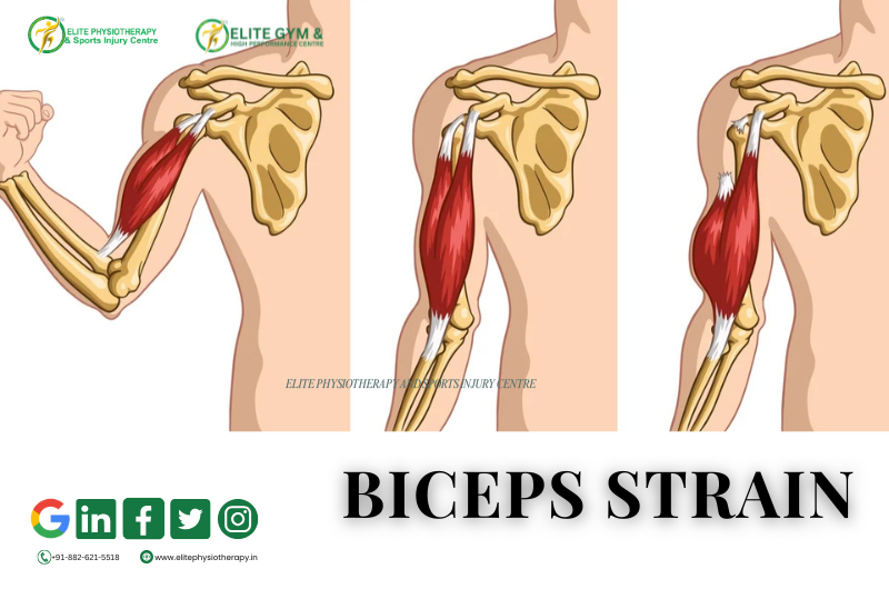
What is a Biceps Strain?

A biceps strain occurs when the biceps brachii muscle or its tendon is torn or overstretched. Usually, a high load or an abrupt force during arm movement causes it. The people most at risk are athletes, weightlifters, and those who lift objects repeatedly.

We frequently see biceps strains in athletes who participate in weightlifting, wrestling, boxing, and racket sports at Elite Physiotherapy and Sports Injury Centre. Early physiotherapy intervention ensures quicker recovery and prevents long-term muscle weakening.

Causes of Biceps Strain

Typically, biceps tension is caused by:

  1. Unexpected eccentric loading when throwing or lifting.
  2. Excessive usage of the arm repeatedly when performing overhead motions.
  3. Direct injury to the upper arm or impact.
  4. Improper lifting technique or improper warm-up.
  5. Loss of control and stability due to muscle exhaustion.

These elements may result in microtears in the tendon or muscle fibers, which would induce discomfort and inflammation.

Classification of Biceps Strain

The degree of muscle fiber damage determines the classification of biceps strains:

  • Grade 1 (Mild): No strength loss, little discomfort, and minimal fiber ripping.
  • Grade 2 (Moderate): Partial tear accompanied by discomfort, edema, and diminished strength.
  • Grade 3 (Severe): Total rupture of a muscle or tendon, frequently accompanied by a protrusion or “Popeye deformity.”

A thorough clinical evaluation at Elite Physiotherapy aids in determining the precise grade and creating a customized rehabilitation strategy.

Clinical Features and Symptoms

Typical signs and symptoms include:

  • The front of the arm hurts sharply.
  • Soreness and swelling above the biceps.
  • Discolouration or bruises.
  • Weakness occurs when the forearms are supinated or the elbows are bent.
  • Tightness in the muscles and trouble lifting things.
  • Obvious deformity brought on by tendon retraction in extreme situations.
  • When the biceps muscle is actively contracted or stretched, pain increases.

Diagnostic Methods and Examination

A thorough patient history and physical examination are the first steps in a clinical diagnosis. To ascertain the injury grade, the physiotherapists at Elite Physiotherapy employ a mix of muscle testing, palpation, and examination.

Special Physiotherapy Tests

  1. Biceps Squeeze Test – Helps detect complete rupture of the distal biceps tendon.
  2. Speed’s Test: Determines whether there is tendinous involvement by measuring pain over the bicipital groove.
  3. Yergason’s Test: Identifies instability in the bicipital groove or pathology in the long head of the biceps tendon.

These tests aid in distinguishing between tendinous damage and muscle belly strain.

Investigations

  • Ultrasound: To detect muscle fiber tears and hematomas.
  • MRI: For severe or chronic cases to assess tendon retraction or complete rupture.

Physiotherapy Management at Elite Physiotherapy and Sports Injury Centre

At Elite Physiotherapy, treatment focuses on pain reduction, restoring strength, and preventing recurrence. Physiotherapists tailor each plan according to the athlete’s sport demands, injury grade, and recovery goals.

Acute Phase (First 3–5 Days):

  • The aim is to reduce pain, inflammation, and swelling through rest, ice, compression, and elevation (RICE) principles. Physiotherapists use advanced modalities such as High-Intensity Laser Therapy (HILT) to accelerate tissue repair and reduce inflammation, while the Super Inductive System (SIS) aids in pain modulation and early muscle activation. Gentle kinesio taping supports the injured muscle and prevents further strain.

Subacute Phase (After 5–10 Days):

  • The focus shifts to restoring flexibility and preventing stiffness. Pain-free active and assisted range of motion exercises are introduced for the elbow and shoulder. Shock Wave Therapy enhances collagen production and promotes tendon healing, whereas Cupping Therapy improves local blood flow and reduces muscle tightness. Physiotherapists apply Dry Needling to deactivate trigger points and reduce muscle tension. Gradual isometric strengthening begins to re-engage the biceps muscle safely.

Strengthening and Functional Phase (After 2–3 Weeks):

  • Once pain subsides, the goal is to rebuild strength and endurance through progressive resistance training, emphasizing eccentric biceps loading. Physiotherapists add functional and sport-specific exercises along with shoulder stabilizer strengthening to improve kinetic chain coordination. The Super Inductive System (SIS) continues to support deep tissue healing and enhance neuromuscular re-education.

Return-to-Sport Phase:

  • The final stage prepares the athlete for competition or full activity. Sport-specific drills, plyometric exercises, and movement retraining are implemented to restore full functional capacity. Physiotherapists conduct postural correction and strength balance assessments to prevent recurrence. Before clearance, the patient must demonstrate full range of motion, pain-free strength, and proper biomechanics.
  • Advanced Modalities Used at Elite Physiotherapy

This combination ensures quicker recovery, reduced pain, and improved performance — helping athletes return to training stronger than before.

Conclusion

If left untreated, a biceps strain can impair arm function and athletic performance. Our state-of-the-art rehabilitation method at Elite Physiotherapy and Sports Injury Centre blends manual treatment with innovative techniques, including Shock Wave, SIS, HILT, Dry Needling, and Cupping.

Athletes who receive individualized care heal more quickly, regain their confidence, and safely resume their best performance.

Triceps Tendinopathy and its Advanced Treatment

What is Triceps Tendinopathy?

A chronic overuse condition of the triceps tendon, usually around or close to its insertion into the olecranon, is known as triceps tendinopathy. It represents degenerative alterations, collagen disarray, and failed tendon repair rather than acute inflammation. Repetitive microtrauma or overloading the tendon can change its structure, causing discomfort and dysfunction.

Tendinopathy is more degenerative than acute tendonitis, which is inflammatory. Particularly in the early stages, it could coexist with certain inflammatory changes, but in chronic situations, tendon degeneration symptoms are typical.

Causes and Risk Factors

Triceps tendinopathy is predisposed by several factors:

  • Weightlifting, throwing, and pushing activities often involve repetitive overload or overuse, which is characterized by repeated extension or resisted loading.
  • Microdamage builds up when training volume or intensity is abruptly increased and adaptation time is inadequate.
  • Poor technique or biomechanics – inadequate shoulder, elbow, or scapular mechanics that cause the tendon to be loaded asymmetrically.
  • Muscle weakness or imbalance: The triceps tendon is subjected to greater strain if stabilizers or synergists are weak.
  • Deficits in nutrition and tendon vascularity: Because tendons have a small blood supply, inadequate microcirculation may hinder repair.
  • Tendon flexibility decreases with age and degenerative changes.
  • Tendon pathology may be exacerbated by comorbidities, which include systemic illnesses such as diabetes, metabolic disorders, or exposure to corticosteroids.

Clinical Features: Signs and Symptoms

When a patient has triceps tendinopathy, they usually exhibit:

  • Gradual development of elbow pain in the back, usually proximal or posterior to the olecranon aspect.
  • Resisted elbow extension or pushing exercises (such as push-ups or bench presses) exacerbate the pain.
  • Palpation of the triceps tendon reveals tenderness, particularly along the tendon or at its insertion.
  • Near the end range of elbow flexion or extension, pain may be triggered.
  • In more severe cases, a slight grating sensation, known as crepitus, may be present.
  • Mild nodules, localized thickening, or swelling of the tendon.
  • suppression of triceps contraction due to weakness or pain.
  • Potential soreness or stiffness following periods of inactivity (e.g., morning stiffness).
  • Decreased functional ability for resistance training or sports in chronic instances.

Physiotherapy Management

Restoring tendon function safely, permitting return to sport, and preventing recurrence are the objectives of an elite sports injury center. Phases can be used to organize the management:

Phase 1: Pain Modulation and Tendon Offloading

  • Activity modification/relative rest: refrain from or reduce activities that cause discomfort (e.g., dips, heavy pressing).
  • Isometric loading – brief, low-to-moderate-intensity isometric holds—helps maintain tendon homeostasis and lessen pain.
  • Cryotherapy, pulsed ultrasonography, and manual soft-tissue treatments (such as tendon massage and cross-friction massage) are modalities for controlling pain and inflammation.
  • Stabilization of the shoulder and scapula ensures that proximal mechanics does not overload the distal tendon.
  • Mobility exercises include maintaining neighboring joint mobility and gently stretching the triceps (within pain tolerance).

Phase 2: Progressive Loading & Remodeling

  • Progressive eccentric loading is the process of gradually increasing load while controlling lowering under resistance.
  • Isotonic concentric loading: start concentric strengthening as soon as the pain is tolerated.
  • Using heavier, slower loading to encourage tendon adaptation is known as heavy slow resistance (HSR) procedures.
  • Integration of sport-specific movement patterns for transfer is known as functional and sport-specific loading.
  • Neuromuscular control: avoid compensatory patterns (shoulder shrugging, trunk movement) and concentrate on the quality of movement.
  • Soft-tissue mobilization and joint mobilization (of the elbow, shoulder, and thoracic spine) are components of adjunctive manual treatment.

3rd Phase: Return to Sport / High Load & Prevention

  • Once the tendon can withstand it, engage in controlled plyometric exercises, such as medicine-ball tosses and explosive push jobs.
  • Return to full training load gradually; keep a close eye on symptoms and make small improvements.
  • Maintaining and strengthening your muscles can help you develop balanced strength in your triceps, biceps, and shoulder stabilizers.
  • Load monitoring and education: make sure you get enough sleep, raise your load gradually, and get technique coaching to prevent recurrence.
  • Patient education, load monitoring, frequent reassessment, and progressive growth are essential components of an exceptional center.

Advanced / Adjunct Modalities:

Advanced techniques, including Shock Wave Therapy, Super Inductive System (SIS), High-Intensity Laser, Dry Needling, and Cupping, are available to you at the premium clinic. Here’s how they could be combined.

  • Shock Wave Therapy (ESWT / radial / focused)
  • In order to promote neovascularization, collagen production, and pain management in chronic tendinopathies, shock wave therapy has been employed.
  • Usually, it is saved for instances that have not improved after six or more months of conservative treatment.
  • Weekly for three to five sessions, protocols typically use low to moderate intensity, with 2,000 to 2,500 impulses per session.
  • Over neurovascular bundles, open wounds, pacemakers, infections, and cancer are among the contraindications.
  • For faster healing, Elite Physiotherapy uses loading protocols in conjunction with shock waves that are carefully focused in the tendon region.
  • Super Inductive System (SIS)
  • SIS, or pulsed electromagnetic induction, is a type of electromagnetic field therapy that is intended to heat tissues deeply and increase cellular activity.
  • The idea is to increase vascularity, lessen pain, and stimulate metabolic activity in degenerative tendon tissue, even if there is little high-level data in triceps tendinopathy specifically.
  • In practice, loading exercises might be preceded with SIS sessions (e.g., 20-30 min) to increase tissue responsiveness and extensibility.
  • High-Intensity Laser Therapy (HILT)
  • Class IV high-intensity lasers can speed up tissue repair, lessen discomfort, and promote photobiomodulation.
  • Laser therapy may improve collagen remodeling, increase mitochondrial function, and decrease inflammation in tendinopathies.
  • Before or after loading workouts, we can administer HILT in cycles (e.g., 2-3 minutes per zone, numerous passes) across the tendon region at the Elite Center, guaranteeing safety and appropriate dosage.
  • Dry Needling and Cupping

Why Elite Physiotherapy & Sports Injury Centre Approach Matters

  • At Elite Physiotherapy, we adhere to an evidence-based, performance-driven methodology:
  • We provide more than simply symptom-based care; we conduct thorough, impartial evaluations.
  • Depending on the athlete’s sport, strength, and tendon capacity, we customize loading progressions.
  • Instead of depending solely on passive treatments, we mix manual and exercise therapy with cutting-edge modalities (SIS, Shock Wave, Laser).
  • In order to prevent flare-ups and guarantee a safe return to sports, we dynamically monitor and modify load.
  • Aware of the athlete’s training requirements, we place a strong emphasis on teaching, biomechanics correction, and recurrence avoidance.
  • Our patients receive top-notch care that goes above and beyond normal rehabilitation.