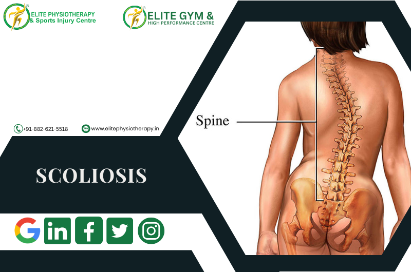
Scoliosis and its management strategies

What Is Scoliosis?

Scoliosis is a three-dimensional spinal deformity characterized by both vertebral rotation (in the axial plane) and lateral curvature of the spine (in the coronal plane). One side of the back may protrude more than the other while bending forward, and the spine may resemble an “S” or “C” when viewed from behind. Scoliosis is typically defined as a curvature of 10 degrees or more, as determined by the Cobb technique.

It is a complicated three-plane problem since sagittal curvatures (kyphosis, lordosis) frequently change beyond the coronal deformity.

In the context of sports injuries and specialist physiotherapy, early identification, thorough evaluation, and customized rehabilitation can aid in managing progression, reducing symptoms, and enhancing function.

Causes & Contributing Factors

Scoliosis arises from multiple possible causes; many cases have an unknown origin. Here are key categories:

1. Idiopathic scoliosis

  • The most prevalent kind, particularly in teenagers (ages 10–18).
  • Subtypes include infantile, juvenile, and adolescent idiopathic.

2. Congenital scoliosis

  • As a result of birth-related vertebral abnormalities (hemivertebra, vertebral fusion).

3. Neuromuscular scoliosis

  • Linked to neuromuscular disorders, such as spinal cord damage, cerebral palsy, and muscular dystrophy.

4. Degenerative (de novo) scoliosis

  • Occurs in adults due to osteoporosis, facet arthropathy, and disc degeneration.

5. Functional (postural, non-structural) scoliosis

  • Adaptable curvature without rotation of the vertebrae.
  • When the underlying cause (muscle spasm, leg-length disparity) is addressed, it goes away.

Types & Classification

To understand and manage scoliosis, clinicians use classifications based on various dimensions:

By Etiology

Idiopathic, congenital, neuromuscular, degenerative, functional (as above)

By Age of Presentation

  • Infantile (< 3 years)
  • Juvenile (4–10 years)
  • Adolescent (> 10 years)

Curve Magnitude (Cobb Angle)

  • Mild: < 20°
  • Moderate: 20°–40° (or 25°–50° in some sources)
  • Severe: > 45°–50°

By Curve Location / Pattern

  • Thoracic curve
  • Thoracolumbar curve
  • Lumbar curve
  • Double / S-shaped curves (two curves)
  • Single or compensatory curves

By Structural Rigidity

  • Structural scoliosis:  Fixed curve with spinal rotation and structural alterations.
  • Non-structural / functional scoliosis: Flexible curvature, no rotation, and reversible by specific motions.

Clinical Features, Signs & Symptoms

While not everyone with scoliosis exhibits symptoms, common clinical characteristics include:

  • Asymmetry in the trunk that is visible (uneven shoulder heights, scapular prominence)
  • Prominence or hump in the ribs when leaning forward
  • Leg-length disparity, pelvic tilt, or uneven waistline
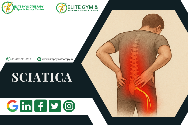
  • Particularly in people with degenerative changes, back pain
  • Stiffness or exhaustion in the paraspinal muscles
  • limited range of motion in the spine
  • Reduced chest expansion and respiratory impairment in severe curvature

Upon physical examination, one could note:

  • Unbalanced paraspinal muscle mass
  • prominent concave side rib hump
  • Trunk shift over the pelvis
  • Variation in the alignment of the spinous process
  • Variations in posture when walking and standing

When asymmetry is detected, young patients frequently show up through school screening (such as Adam’s forward-bend test).

Physical Tests & Assessment Techniques

Assess paraspinal muscle tightness or tenderness.

Special Orthopedic Tests

1. Adam’s Test of the Forward Bend

Examiners look for rib protrusion or asymmetry from behind.

2. Inclinometer/Scoliometer

When bending forward, the angle of trunk rotation, or ATR, is measured.

helps measure the prominence of the rib hump.

3. Landmarks of the spine and palpation

To find rotation or lateral deviation, feel for spinous processes.

4. Testing for Range of Motion (ROM)

trunk rotation, flexion/extension, and lateral flexion

Compare the flexibility from side to side.

5. Measurement of Leg Length

To eliminate the possibility that a leg-length disparity is causing a functional curve

Assess the curve’s flexibility using the side-bending test to see if it bends correctly. Torsional stress tests and the difference between the ribs and vertebrae. Provocative testing for disc, facet, and nerve root involvement; segmental vertebral mobility tests (passive intervertebral motion)

Physiotherapy Management Strategies

At Elite Physiotherapy & Sports Injury Centre, we emphasize evidence-based, patient-centred interventions. Our goal is to slow or halt curve progression, reduce pain, restore alignment and mobility, and enhance function.

1. Physiotherapeutic Scoliosis-Specific Exercises (PSSE / 3D Correction)

These specific remedial workouts are made to fit the curvature pattern of each patient. Among the principles are:

  • Self-correction in three dimensions: Active correction in the transverse, sagittal, and coronal planes
  • Stabilization of the posture correction
  • Breathing exercises to de-rotate the rib cage and extend the concave side
  • Integration with ADLs (training for habitual posture)
  • Balance, core activation, and sensorimotor training

2. Manual Therapy & Mobilization

  • Gentle spinal mobilization of restricted segments
  • Myofascial release and soft tissue mobilization of tense muscles on the convex side
  • Increasing thoracic mobility using rib mobilization
  • Elongated muscle strengthening and shortened muscle stretching

3. Core Stabilization & Motor Control Training

  • Activation of deep trunk muscles (transverse abdominis, multifidus)
  • During movement, functional trunk control
  • Training for balance and proprioception

4. Electro-physical Modalities

Through pain relief, soft tissue modulation, and improved muscle function, these modalities can be used in conjunction with active and manual approaches. Each modality may be integrated as follows:

5. Bracing / Orthosis (in collaboration with orthopedics)

  • When indicated (moderate curves in growing patients), physiotherapy works alongside bracing:
  • Teach patients how to perform corrective exercises within the brace
  • Postural coaching with brace off
  • Strengthening to manage brace-induced weakness

Why Patients Should Choose Elite Physiotherapy & Sports Injury Centre

At Elite, we bring together:

  • proficiency in spinal and musculoskeletal care
  • Personalized procedures for scoliosis
  • Availability of auxiliary cutting-edge modalities such as high-intensity laser, shock wave, and Super Inductive System
  • Prioritize functional rehabilitation so you can resume your daily activities or sports.
  • Thorough monitoring, follow-up, and high-quality results

Conclusion

Being a complicated, three-dimensional abnormality, scoliosis necessitates meticulous evaluation, specialized treatment, and regular monitoring. At Elite Physiotherapy & Sports Injury Centre, we provide patients with a comprehensive, evidence-based treatment plan that includes everything from functional reintegration and long-term monitoring to precise orthopedic physical assessment, curve-specific corrective techniques, supportive modalities like shock wave, Super Inductive system, high-intensity laser, and selective dry needling or cupping.

We seek to control curve progression, reduce discomfort, improve mobility, and assist you in leading an active, satisfying life by emphasizing individualized rehabilitation.

Ankylosing Spondylitis : Back Pain

A chronic inflammatory disease that mostly affects the axial skeleton, ankylosing spondylitis (AS) can cause pain, stiffness, and even spinal fusion. To effectively manage AS, we at Elite Physiotherapy and Sports Injury Centre provide individualized, evidence-based physiotherapy interventions.

Understanding Ankylosing Spondylitis

The hallmark of ankylosing spondylitis, a form of axial spondyloarthritis, is inflammation in the spine and sacroiliac joints. Parts of the spine may fuse as a result of the new bone that forms as a result of this inflammation over time. Although the precise reason is yet unknown, genetic factors—specifically, the presence of the HLA-B27 gene—have a significant impact. Environmental variables may also influence the start and progression of disease.

Causes and Classification

AS is under the category of inflammatory rheumatic diseases known as spondyloarthritis. An autoimmune reaction, in which the body’s immune system targets its own tissues, is thought to be the main cause. One known risk factor is genetic predisposition, particularly the HLA-B27 gene. Infections and other environmental causes may also contribute to the development of disease.

Clinical Features and Symptoms

Common symptoms of AS include:

  • Persistent stiffness and discomfort in the lower back, particularly in the morning or after extended periods of inactivity.
  • Peripheral joints such as the shoulders, knees, and hips may experience pain and swelling.
  • Inflammation where tendons and ligaments connect to bones is called enthesitis.
  • Fatigue and decreased mobility.
  • A stiff spine may result from spinal fusion in more advanced stages.

Diagnostic Methods

Diagnosis of AS involves:

  • Clinical Evaluation: Evaluation of medical history and symptoms.
  • Imaging: MRI or X-rays to identify alterations in the spine and sacroiliac joints.
  • Laboratory Tests: Blood tests to look for the HLA-B27 gene and indicators of inflammation.

Physiotherapy Management at Elite Physiotherapy and Sports Injury Centre

At Elite Physiotherapy and Sports Injury Centre, we provide a comprehensive physiotherapy approach tailored to the needs of AS patients:

1. Assessment Techniques

  • A comprehensive evaluation is carried out utilizing:
  • Postural Analysis: Finding any anomalies or compensatory patterns.
  • Range of Motion (ROM) Measurements:  Evaluating joint mobility and flexibility.
  • Strength Testing: It is the process of assessing the endurance and strength of muscles.
  • Special Tests: To evaluate spinal and sacroiliac joint involvement, particular orthopedic tests are used. such as the Schober test, the SI compression test, and Gaenslen’s test, etc.

2. Treatment Modalities

To improve therapeutic results, we use cutting-edge modalities:

  • Shock Wave Therapy: Using acoustic waves, shock wave therapy helps musculoskeletal tissues repair.
  • Super Inductive System: Reduces pain and inflammation by stimulating deep tissues with high-intensity electromagnetic fields.
  • High-Intensity Laser Therapy: Reduces inflammation and speeds up tissue repair by using concentrated light.
  • Dry Needling and Cupping Therapy: Uses specific procedures to relieve pain and tightness in the muscles.

3. Exercise Therapy

A personalized exercise program focusing on:

  • Exercises for Spinal Mobility: To preserve and enhance flexibility.
  • Strengthening exercises: These support spinal alignment by focusing on the postural and core muscles.
  • Aerobic Conditioning: To lessen tiredness and increase general fitness.

Why Choose Elite Physiotherapy and Sports Injury Centre?

Our center offers:

  1. Expert Physiotherapists: Specialized in managing complex conditions like AS.
  2. State-of-the-Art Equipment: Utilizing advanced therapeutic modalities.
  3. Holistic Approach: Addressing physical, emotional, and lifestyle aspects of AS.
  4. Personalized Care: Tailored treatment plans to meet individual needs.

If you’re experiencing symptoms of Ankylosing Spondylitis or seeking specialized care, contact Elite Physiotherapy and Sports Injury Centre today. Our dedicated team is here to support you on your journey to better health and mobility.

Sciatica – Culprit for your long running lower back pain

A common condition that causes pain, numbness, or weakness along the sciatic nerve is sciatica. This nerve travels down each leg from the lower back through the buttocks. Our goal at Elite Physiotherapy and Sports Injury Centre is to improve function, reduce pain, and enhance quality of life through individualized treatment strategies.

What is Sciatica?

Pain that travels along the sciatic nerve route is referred to as sciatica. Usually, it impacts one side of the body. The pain might hamper everyday activities and ranges from mild to severe. Even while it usually goes away on its own, persistent cases need professional attention.

Causes of Sciatica

  • Herniated Disc: The nerve root is compressed by a slipped disc.
  • Spinal Stenosis: The nerve is compressed when the spinal canal narrows.
  • Degenerative Disc Disease: Nerve discomfort is caused by disc wear.
  • Spondylolisthesis: A nerve is pinched when one vertebra slides across another.
  • Piriformis Syndrome: The piriformis muscle irritates the sciatic nerve.
  • Trauma or Injury: Nerve damage can result from accidents.
  • Infections or tumors are uncommon but possible causes.

Clinical Features, Signs, and Symptoms

  • Sharp, shooting pain from the lower back to leg.
  • Tingling or numbness in the foot or leg.
  • Weakening of the leg muscles.
  • Prolonged standing or sitting causes the pain to worsen.
  • walking or carrying out regular tasks with difficulty.

Diagnostic Methods and Special Tests

An accurate diagnosis is essential for successful treatment. We conduct a comprehensive evaluation at Elite Physiotherapy and Sports Injury Centre by integrating a physical examination and patient history.

Common Diagnostic Techniques:

  • The Straight Leg Raise (SLR) test: Involves raising the straightened leg to assess nerve root irritation. Pain reproduction suggests sciatica.
  • Slump Test: The patient sits with their knee extended and their spine flexed. If sciatic symptoms show up, it’s positive.
  • Crossed Straight Leg Raise Test: Lifting the unaffected leg while replicating symptoms indicates a disc herniation.
  • Neurological Examination: Assesses strength, reflexes, and sensory abilities.
  • Doctors may recommend imaging tests such as MRIs or CT scans to confirm disc herniation or stenosis.

Physiotherapy Management of Sciatica

At Elite Physiotherapy, we aim to reduce discomfort, increase range of motion, and stop recurrence. We emphasize individualized, evidence-based strategies with premium modalities.

1. Manual Therapy

  • To increase spinal mobility and lower nerve pressure, we use soft tissue methods and joint mobilization.

2. Therapeutic Exercises

  • Stretching: To relieve tension, focus on the piriformis and hamstrings.
  • Exercises for Stabilization: Use low-load core strengthening to enhance spinal support.
  • Exercises for Nerve Mobilization: Encourage sciatic nerve mobility without making symptoms worse.

3. High-End Modalities

A. Shock Wave Therapy

  • Efficient in promoting blood flow and nerve regeneration to lessen chronic pain.

B. Super Inductive System (SIS)

  • Provides deep electromagnetic pulses to the afflicted area to increase circulation and decrease inflammation.

C. High-Intensity Laser Therapy

  • Reduces inflammation, discomfort, and promotes healing by penetrating deep tissues.

D. Dry Needling & Cupping Therapy

  • Physiotherapists target trigger points and relieve tense muscles, which is especially beneficial for sciatica caused by piriformis syndrome.

4. Postural Correction and Ergonomic Exercises

  • We provide home home-based exercise program for your posture correction that will help to minimize nerve compression.

Why Choose Elite Physiotherapy and Sports Injury Centre?

For sciatica, we offer a comprehensive, patient-focused therapy. Our skilled therapists and cutting-edge methods guarantee a quicker, secure recovery. We ensure long-term relief by tailoring each plan to each client’s unique demands.

At Elite Physiotherapy, we think that in order to assist patients properly heal from sciatica, we need to combine science, technology, and compassionate treatment.

Piriformis Syndrome and its Elite Management

What is Piriformis Syndrome?

Behind the gluteus maximus, deep in the buttocks, is a tiny, flat muscle called the piriformis. It joins the apex of the femur to the sacrum. Its main function is to support the pelvis and aid in hip rotation when walking or running. The sciatic nerve that passes through or beneath the piriformis muscle may be compressed if it gets tense, swollen, or spasms. The result of this is Piriformis Syndrome.

Causes of Piriformis Syndrome

  • Overuse of muscles from repetitive sports like cycling or jogging
  • Damage or trauma to the gluteal area
  • Extended periods of sitting, particularly with bad posture
  • Variations in anatomy, where the piriformis is where the sciatic nerve travels
  • Piriformis muscle inflammation or spasm
  • Conditions affecting the lumbar spine that lead to transferred sciatic pain

Classification of Piriformis Syndrome

Although there is no strict classification system, it is commonly categorized based on the cause:

  • Primary Piriformis Syndrome: brought on by congenital defects or anatomical variances.
  • Secondary Piriformis Syndrome: Caused by inflammation, misuse of the muscles, or trauma.

Clinical Features / Signs & Symptoms

  • An excruciating, deep buttock pain
  • Often misdiagnosed as sciatica, pain radiating down the back of the thigh
  • Along the sciatic nerve distribution, numbness and tingling
  • Increased discomfort when sitting or moving the hips
  • The piriformis muscle is painful.
  • Having trouble walking or climbing stairs, for example

Diagnostic Methods, Tests, and Physical Examination

Physical Examination and Special Tests

  • Palpation: Potential involvement is indicated by tenderness over the piriformis muscle.
  • Piriformis Muscle Stretch Test: The patient is in a supine position while the examiner passively rotates, adducts, and flexes the hip. Piriformis involvement is suggested by pain reproduction.
  • Freiberg Test: Pain occurs when the extended thigh is forced to rotate internally.
  • Pace Test: Pain in the piriformis region is caused by outward rotation of the hip and resisted abduction.
  • Straight Leg Raise Test: It may be positive in certain situations; however, it aids in ruling out lumbar disc herniation.
  • Imaging and Other Investigations: MRI or Ultrasound Imaging

Physiotherapy Management at Elite Physiotherapy and Sports Injury Centre

1. Manual Therapy and Soft Tissue Techniques

  • Piriformis Muscle Release: Expert soft tissue release eases sciatic nerve constriction and eases tense muscles.
  • Trigger point therapy: It reduces pain and spasms by applying direct pressure to muscular knots.

2. Therapeutic Exercises

  • Stretching exercises that target the hamstrings, hip adductors, and piriformis to release tension.
  • Strengthening exercises: To increase pelvic stability, strengthen your gluteus medius and core muscles.
  • Exercises for Postural Correction: Focusing on spinal alignment and good sitting posture.

3. High-End Physiotherapy Modalities

  • At Elite Physiotherapy and Sports Injury Centre, we employ advanced modalities for effective treatment:
  • Shock Wave Therapy (SWT): High-energy waves stimulate tissue repair, reduce inflammation, and improve circulation.
  • Super Inductive System (SIS): Provides deep heating, enhancing muscle relaxation and reducing spasms.
  • High Intensity Laser Therapy (HILT): Accelerates tissue healing and reduces nerve-related pain by penetrating deep tissues.
  • Dry Needling: Thin needles target trigger points to release muscle tension and improve blood flow.
  • Cupping Therapy: Increases circulation and releases fascial restrictions in the piriformis and surrounding tissues.

4. Education and Lifestyle Modification

  • Educating patients on avoiding prolonged sitting, proper stretching, and regular exercise prevents recurrence.

5. Home Exercise Program

  • We provide personalized home exercise programs, including stretching and strengthening exercises, to maintain progress.

Why Choose Elite Physiotherapy and Sports Injury Centre?

At Elite Physiotherapy and Sports Injury Centre, we provide highly individualized physiotherapy service together with cutting-edge diagnostic methods. Utilizing cutting-edge technologies and evidence-based techniques, our skilled team specializes in treating Piriformis Syndrome. We guarantee that each patient gets a comprehensive evaluation and a customized treatment strategy.

Combining manual therapy, therapeutic exercises, and state-of-the-art techniques like Shock Wave, SIS, HILT, Dry Needling, and Cupping improves our outcomes. We provide patients with the information and resources they need for long-term recovery in addition to treatment.

Understand Spondylolisthesis

What is Spondylolisthesis?

The sliding of one vertebral body relative to the next, known as spondylolisthesis, results in pain, radicular symptoms, or mechanical symptoms. It may result from idiopathic, acquired, or congenital causes. The degree of slippage (Meyerding Classification) of one vertebral body on the next vertebral body determines the grade of spondylolisthesis.[1]

What Causes Spondylolisthesis?

  • Congenital weakness, stress fractures, and spinal degeneration are common reasons.
  • Athletes’ recurrent stress or aging-related deterioration may potentially be the cause.
  • Traumatic accidental injuries
  • Pathological factors, such as connective tissue disorders and neoplasms.
  • Following a laminectomy.
  • Early-life decline in neural arch strength

Clinical Presentation

Pain presentation

  • Low back pain (lumbar) or neck pain (cervical)
  • Can mimic radiculopathy
  • Worsens with spinal extension or palpation
  • Improves in flexed or supine posture

Neurological involvement

  • Radicular pain due to nerve root compression (foraminal narrowing, disc protrusion, or canal stenosis)
  • Reduced spinal ROM
  • Possible neurological deficits

Musculoskeletal findings

  • Hamstring tightness or spasm
  • Muscle weakness and atrophy
  • Crouched gait (Phalen-Dickson sign) due to postural compensations

Functional impairments

  • Disturbed coordination and balance
  • Difficulty walking

Elite Physiotherapy’s Personalized Approach

At Elite Physiotherapy & Sports Injury Centre, we value your unique needs. We start with careful evaluation. Design a plan that fits your lifestyle and pain level. We blend manual therapy, therapeutic exercises, and advanced modalities to restore function and strength.

Assessment Technique & Specialized Physiotherapy Test

To fully assess spondylolisthesis, we use a multi-step approach:

Postural Analysis

  • Observe spinal alignment, pelvic tilt, and posture during standing and movement.
  • Look for abnormal lumbar curves or forward displacement.

Movement Testing

  • Test active and passive spine flexion, extension, and side-bending.
  • Note pain levels and movement restriction.

Specific Segmental Move Test

  • Applying targeted mobilization to each lumbar segment while feeling for resistance or movement.
  • This reveals the precise vertebra level that has shifted or lacks stability.

Functional Load Assessment

  • Observe how the spine responds under gentle compressive load in neutral or extension.
  • Assess pain response and stability under real-life stress.

Neurological Screen (if needed)

  • Check reflexes, muscle strength, and sensation in lower limbs.
  • Rule out nerve compression from the shifted vertebra.

Personalized Physiotherapy Management

1. Pain Relief and Initial Stabilization

  • Begin with gentle manual therapy targeting the affected vertebral level.
  • Use soft-tissue release to ease muscle tightness.
  • Apply electrotherapy (like TENS or ultrasound) to relieve pain and reduce muscle spasm.

2. Core Stabilization and Neutral Spine Control

  • Teach deep abdominal bracing in neutral spine position.
  • Guide pelvic tilts and glut activation exercises to reduce spinal load.
  • Progress into bird-dog, focusing on spine control with limb movement.

3. Lumbar Segmental Mobilization

  • Apply controlled mobilization at the specific slipping segment.
  • Use directional preference movements to encourage vertebral alignment.
  • Reassess after each session to ensure progress.

4. Flexibility and Paraspinal Strengthening

  • Stretch tight hamstring and hip flexor muscles without forcing movement.
  • Incorporate side-planks and modified bridges to strengthen trunk and glutes.
  • Progress with Swiss-ball control exercises for added stability.

5. Functional Training and Movement Re-education

  • Advise and train safe movement patterns for daily tasks.
  • Use squat-to-stand drills with neutral spine emphasis.
  • Incorporate dynamic balance work, such as single-leg stance with trunk rotation.

6. Gradual Return to Sport or Activities

  • Include sport-specific drills with controlled spine posture.
  • Gradually increase intensity, speed, and complexity.
  • Monitor form to prevent shear forces on the lumbar spine.

7. Education and Prevention

  • Teach proper posture and body mechanics for activities such as bending, lifting, and sitting.
  • Provide a personalized home exercise program for spinal stability.
  • Schedule periodic follow-ups to track progress and adjust treatment.

Why Elite Physiotherapy Excels

  • We offer expert, hands-on care tailored to each slip’s severity.
  • We maintain a safe, progressive plan built on spinal mechanics.
  • We combine manual therapy, movement control, and sport readiness.
  • We stress patient education, empowering long-term spine health.
  • We monitor closely and adapt therapy as you improve.

Conclusion

At Elite Physiotherapy & Sports Injury Centre, we guide healing from spondylolisthesis with precision, compassion, and progression. We blend assessment with active treatment, core retraining, and safe return to activity. We help you reclaim pain-free movement with strength, control, and confidence.

Cross Bracing Protocol (CBP) for ACL Rehab

What is the Cross Bracing Protocol?

For certain acute ACL ruptures, the Cross Bracing Protocol (CBP) is a time-sensitive, non-surgical treatment option. For the first four weeks, the knee is in a 90° flexion position. After that, the brace is gradually unlocked to restore motion, to mimic the remaining ACL so that the native ligament can repair. Early results from prospective cohorts report high rates of MRI continuity at 3 months; clinical trials are still being conducted to determine suitable individuals and longer-term outcomes.

Why it matters

A totally ruptured ACL was formerly thought to be incapable of healing, necessitating early repair or conventional rehabilitation without bracing. Recent data casts doubt on that belief. In a secondary analysis of the KANON trial, approximately one-third of ACLs treated solely with rehabilitation had MRI evidence of healing at two years (and roughly one-half when those who underwent surgery were excluded), and the healed group had better patient-reported outcomes. By purposefully placing the knee to encourage tissue apposition as soon as possible after injury, CBP enhances this healing potential.

Who is (and isn’t) a Candidate?

The best candidates typically show up early (preferably within 10 days after the injury), have an MRI showing an acute ACL rupture, and can follow bracing and follow-up instructions to the letter. MRI morphology (such as remnant quality, displacement, and gap distance) and patient characteristics (sport demands, support, and comorbidities) are progressively taken into account during the selection process.

Not suitable for all: Clinicians are frequently pushed onto alternative approaches due to multi-ligament injuries, displaced bucket-handle meniscal tears/loose bodies requiring urgent surgery, extremely delayed presentation, or enhanced thrombosis risk. (CBP-using programs typically sort them out in the early stages of MRI-guided decision making.)

How the Protocol Works

The basic idea is to protect the healing ACL from anterior tibial translation and pivoting by initially reducing and immobilizing (similar to a fracture) and then gradually restoring motion every week.

Weeks 1–4

  • Brace: locked at 90° (24/7; sleep in brace).
  • Weight-bearing: Non-weight-bearing (NWB) with crutches.
  • Goals: protect the healing zone, maintain patellofemoral mobility, prevent deconditioning (safe isometrics).

5th Week

  • Brace 60–90°. Continue NWB. Begin gentle, brace-permitted ROM drills; progress isometric hamstring/quadriceps sets in allowed angles.

6th Week

  • Brace ~45–90°. Still NWB. Add stationary bike within brace limits if permitted.

7th Week

  • Brace 30–120°. Partial weight-bearing begins; gait retraining within brace range. Light closed-chain work in safe angles.

Week 8

  • Brace 20–120°. Progress PWB loading, proprioception in brace.

Week 9

  • Brace 10–120°. Full weight-bearing as tolerated in brace; advance strength, balance, conditioning tasks (pool if available) without pivoting/cutting.

Weeks 10–11

  • Unrestricted brace during the day; remove for sleep. Continue progressive strengthening, linear conditioning, and landing mechanics in straight plane.

Week 12

  • MRI and clinical review. The brace is taken off, and criterion-based rehabilitation continues if the MRI reveals sufficient continuity and clinical stability. If not, a “cross-over” to surgery or an extension of bracing may be part of the collaborative decision-making process.

Beyond 12 weeks (typical milestones)

  • Weeks 16–17: Start straight-line running (criteria-based).
  • ~6–12 months: Athletes gradually return to training and then progress to sport-specific change of direction once they achieve goals for strength, symmetry, hop testing, and movement quality. Many programs plan for nine to twelve months before a complete return to pivoting sports.

Rehabilitation Priorities

Protection & monitoring (0–12 wks)

  • Teach people to use crutches, wear braces strictly, and refrain from twisting or pivoting.
  • Ankle pumps, hip/glute/hamstring isometrics, and early patellar mobilization (knee maintained within brace limits).
  • Some programs aggressively monitor DVT risk (local techniques vary; some reported early DVTs spurred teams to embrace pharmacologic prophylaxis).

Strength & motor control (weeks 5–12)

  • Avoid anterior shear (no open-chain knee extension in vulnerable ranges) and increase closed-chain strength in safe arcs.
  • Brace ROM connects directly with balance, trunk/hip control, and graded conditioning (bike, pool, and later treadmill).

Run-jump-cut reconditioning (post-brace)

  • Linear running → decel/accel → low-level plyometrics → planned change-of-direction → unplanned COD and sport skills after meeting patient-reported outcomes targets and ROM/strength/hop/movement benchmarks.

To prevent vasoconstriction during the initial healing window, some CBP teams prohibit the use of NSAIDs, knee aspiration, and even cryotherapy.

What Does the Evidence Say (so far)?

  • High MRI early healing with CBP: Three months after CBP, a prospective cohort reported ~90% ACL continuity, and patients with greater early MRI healing achieved better results. Researchers still need randomized longer-term results.
  • In certain situations, ACLs can heal without surgery: In the KANON dataset, MRI showed healing in around 30 to 50% of patients who underwent rehabilitation alone, independent of CBP.. The KOOS results of the healed groups were better than those of the non-healed/reconstructed groups.
  • Risk-benefit balance: Although reviews point to encouraging healing, they also emphasize the danger of stiffness, the necessity of careful selection and adherence, and the possibility that some patients will require surgery.

CBP vs. Traditional ACL Management

DimensionCross Bracing ProtocolTraditional Reconstruction / Standard Non-Op
Primary goalHeal the native ACL (biologic healing)Replace with graft (surgery) or compensate via neuromuscular rehab
Early positioningImmobilize at 90° flexion for 4 weeks, then staged ROMPrioritize full extension early; brace often locked in extension initially post-op; no prolonged flexion immobilization
Weight-bearing (early)NWB first weeks; add load as brace range increasesWBAT early after ACLR; progress as swelling/quad control allow
ROM strategyDelayed extension; weekly unlock scheduleImmediate mobilization, especially regain full extension to avoid arthrofibrosis
Cryotherapy & NSAIDsSome programs limit early NSAIDs/icing (program-specific)Commonly used to control pain/effusion post-injury/surgery
MonitoringScheduled MRI at ~3 months to confirm continuityImaging usually not required once post-op course is stable
Time to pivot sportsTypically ≥9–12 months and criteria-basedAlso ≥9–12 months and criteria-based (graft maturity & testing)
Key risksStiffness/extension loss if mishandled; non-healing → cross-over to surgeryGraft failure, donor-site morbidity, cyclops lesions, and surgical risks
Time to pivot sportsMotivated, early-presenting patients willing to adhere strictly; favorable MRI patternGraft failure, donor-site morbidity, cyclops lesions, surgical risks

At Elite Physiotherapy and Sports Injury Centre, we integrate the latest evidence-based approaches, such as the Cross-Bracing Protocol (CBP), alongside traditional ACL rehabilitation and surgical recovery programs. With our advanced facilities—including Super Inductive Stimulation (SIS), Class 4 Laser Therapy, CRET Therapy, Shockwave Therapy, and Hydrotherapy—we ensure that every patient receives a personalized, high-end rehabilitation plan. Our focus is not just on healing the ligament but also on restoring strength, balance, and performance, so athletes and active individuals can safely return to their sport or lifestyle with confidence.