Athletes who engage in frequent overhead or throwing motions are susceptible to ulnar collateral ligament (UCL sprain ). The Lite Physiotherapy and Sports Injury Centre offers cutting-edge, research-based treatment for this distressing condition. To restore strength, stability, and performance, the clinic employs cutting-edge modalities and contemporary assessment techniques.
What Is a UCL Sprain?
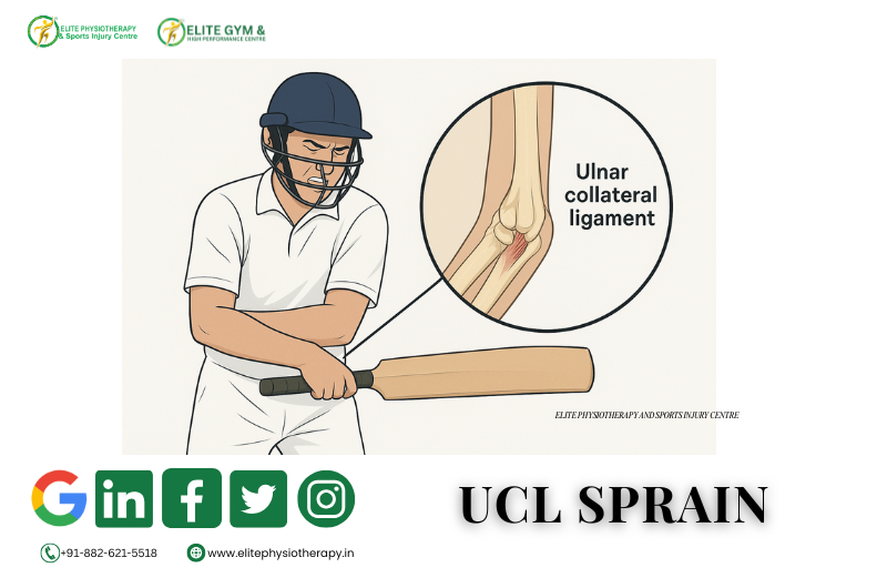
A partial or total rupture of the ligament on the inside of the elbow is known as a UCL sprain. During valgus stress, this ligament keeps the elbow stable. Pain and instability arise when the ligament tears or overstretches. Throwers, wrestlers, weightlifters, Ballers in cricket and athletes participating in racquet sports are frequently affected by the injury.
Causes of UCL Sprain
- Throwing repeatedly puts stress on the medial elbow.
- Poor throwing mechanics increase the valgus load.
- The ligament is strained by abrupt falls or direct impact.
- Dynamic stability is diminished by muscle weakness.
- Micro-trauma accumulates as a result of overtraining.
To guarantee precise treatment planning, we at Elite Physiotherapy pinpoint the precise cause.
Types or Classification of UCL Sprains
- Grade I
- The ligament is overstretched but intact. Pain is present but stability is maintained.
- Grade II
- A partial tear occurs. Pain increases and mild instability appears.
- Grade III
- A complete tear occurs. Instability is obvious during movement or loading.
Proper diagnosis is essential because management differs for each grade.
Clinical Features / Signs and Symptoms
- Medial elbow pain during throwing
- Pain during resisted wrist flexion
- Tenderness along the UCL
- Decreased throwing speed or power
- A feeling of looseness in the elbow
- Swelling around the medial joint line
- Pain during valgus stress
Diagnostic Methods and Investigations
- Clinical Examination
- A thorough history pinpoints the injury mechanism, exercise volume, and symptoms.
Special Tests
- Valgus Stress Test
- In this test, the elbow is subjected to a controlled valgus force at 20 to 30 degrees. A UCL damage is indicated by increased pain or severe gapping.
- Moving Valgus Stress Test
- The examiner applies valgus stress while moving the elbow from full flexion to extension. UCL pathology is indicated by pain that ranges from 70 to 120 degrees.
- Milking maneuver
- To produce a valgus force, the clinician tugs the patient’s thumb. UCL involvement is suggested by medial discomfort.
Without citing any outside sources, these tests aid in confirming the diagnosis.
Imaging
- Ultrasound, MRI, X-ray
Physiotherapy Management of UCL Sprain at Elite Physiotherapy and Sports Injury Centre
- Physiotherapy remains the first-line treatment for most UCL injuries. At Elite Physiotherapy, we uses advanced techniques and high-end modalities to ensure fast and safe recovery.
1. Pain Reduction and Inflammation Control
- Cryotherapy
- During the acute period, cold treatment lessens inflammation.
- Shock Wave Treatment
- Shock wave therapy lessens persistent discomfort and encourages tissue regeneration.
- High-Power Laser Treatment
- This technique enhances collagen alignment and speeds up recovery.
- System Super Inductive (SIS)
- SIS rapidly lessens pain by promoting deep muscle activity.
- Dry Needling
- Dry needling lessens medial elbow strain and relaxes tense muscle bands.
- Cupping Treatment
- Cupping lessens fascial limitations around the elbow and increases blood flow.
- These techniques promote quicker recovery and less pain.
2. Restoring Range of Motion
- Elbow stiffness is lessened, and elbow movement is restored with gentle mobility exercises.
- Flexion and extension of the elbow passively
- Pronation and supination of the forearm
- Drills for shoulder mobility to lessen compensatory tightness
- Adhesive tissue development is inhibited by controlled movement.
3. Strengthening Program
- Elite Physiotherapy employs a methodical approach to strengthening.
- Phase 1: Strengthening Isometrically
- Wrist flexor, extensor, and pronator pain-free isometrics start early.
- Phase 2: Strengthening Dynamically
- Light resistance bands strengthen the flexor-pronator mass.
- Scapular stability exercises enhance shoulder mechanics.
- Eccentric loading enhances tendon resilience.
- Phase 3: Advanced Strengthening
- Eccentric loading enhances tendon resilience.
- Plyometric exercises prime the elbow for high-intensity sports.
4. Correction of Throwing Mechanics
- The corrective procedure prevents recurrence.
- Elite Physiotherapy examines throwing patterns and makes the following corrections:
- Position of the arm slot
- Mechanisms of trunk rotation
- Pattern of follow-through
- Distribution of force among joints
- The corrective procedure prevents recurrence.
5. Proprioception and Neuromuscular Training
- Proprioceptive exercises improve dynamic elbow stability.
- Drills using weight-bearing
- Ball-catch exercises
- Techniques for functional tapping
- These workouts get the athlete ready for duties unique to their sport.
6. Return-to-Sport Programming
- A methodical return procedure ensures safety.
- Stage 1: Regulated Throwing
- Athletes use low volume for light tosses.
- Stage 2: Throwing Progressively
- While keeping an eye on symptoms, intensity progressively rises.
- Stage 3: Exercises Particular to Sports
- Athletes restart competitive or fast-paced drills.
- Physiotherapists at Elite Physiotherapy customize this path for every athlete.
7. Elite Physiotherapy’s Preventive Techniques
- For long-term elbow health, prevention is crucial.
- Frequent evaluations of strength
- Monitoring the throwing load
- Maintaining mobility
- Soft tissue maintenance
- Corrections for skill-based training
- These methods ensure long-term performance and safety..
Conclusion
- Although dangerous, a UCL sprain is treatable. Athletes can safely return to sport with the right diagnosis, cutting-edge physiotherapy, and contemporary techniques. At Elite Physiotherapy and Sports Injury Centre, we use cutting-edge technologies like Shock Wave, SIS, High-Intensity Laser, Dry Needling, and Cupping to provide individualized, evidence-based rehabilitation. Our methodical evaluation and rehabilitation process ensures long-term elbow stability and full recovery.