LISFRANC INJURY OF THE FOOT

Introduction

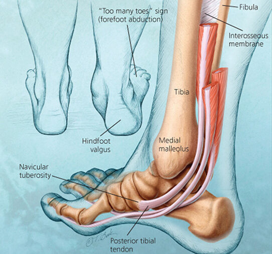
A Lisfranc injury affects the midfoot, involving the tarsometatarsal joints and associated ligaments. A prompt and precise diagnosis is essential for successful treatment and the best possible outcome. Each patient receives individualized care at Elite Physiotherapy and Sports Injury Centre, where we specialize in cutting-edge physiotherapy therapies designed to address Lisfranc issues. ​

Causes and Mechanism of Injury

Lisfranc injuries can result from both direct and indirect trauma:​

  • Direct Trauma: A force exerted to the top of the foot, as in car accidents, crush injuries, or falls from a height, is frequently the cause of direct trauma. ​
  • Indirect Trauma: Twisting on a plantarly flexed foot or axial loading through the foot are common causes of indirect trauma, which is commonly observed in sports like football or horseback riding. ​

Signs and Symptoms

Patients with a Lisfranc injury may exhibit:​

  • Pain and swelling in the Midfoot.​
  • Bruising on the bottom of the foot.​
  • Pain when bearing weight, particularly when going downstairs.​
  • Tenderness when the midfoot region is touched.​
  • Difficulty or inability to bear weight on the affected foot. ​

Diagnostic Methods

Accurate diagnosis involves a combination of clinical assessment and imaging studies:​

  • Clinical Examination: Key assessments include:​
    • keeping an eye out for bruises and swelling, especially on the bottom of the foot.​
    • Palpating the midfoot to identify areas of tenderness.​
  • Performing specific tests, such as:​
    • Piano-Key Test: Moving the metatarsals up and down to detect pain or instability.
    • Midfoot Compression Test: Squeezing the foot’s width to stress the space between the first and second metatarsals, noting any pain or clicking sounds.
    • Passive Pronation-Abduction Test: Rotating the forefoot outward while stabilizing the hindfoot to assess for pain, indicating potential injury.
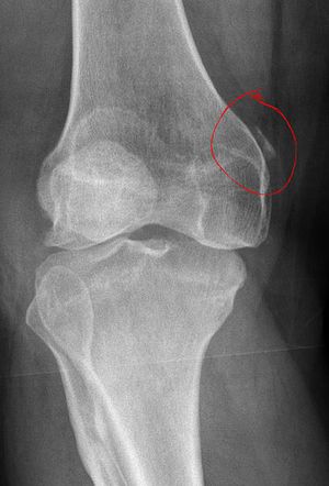
  • Imaging Studies:
    • X-Rays: Weight-bearing X-rays are essential to reveal joint misalignments or fractures.​
    • CT Scans: Useful for detecting subtle fractures and planning surgical interventions.​
    • MRI: Effective in evaluating ligament injuries and assessing soft tissue damage. ​

Physiotherapy Management at Elite Physiotherapy and Sports Injury Centre

At Elite Physiotherapy and Sports Injury Centre, we offer a comprehensive physiotherapy program for Lisfranc injuries, focusing on:​

  • Edema Reduction: Utilizing techniques like cryotherapy to minimize swelling and promote healing.​
  • Strengthening Exercises: Addressing muscle weakness resulting from immobilization through targeted exercises.​
    • Toe Flexor and Extensor Strengthening
    • Ankle and Foot Isometric Strengthening
    • Resistance Band Exercises
    • Heel and Arch Strengthening
    • Balance and Proprioception Training
    • Functional and Sport-Specific Strengthening
  • Flexibility Training: Improving range of motion in the foot and ankle to restore standard movement patterns.​
    • Toe and Forefoot Flexibility Exercises
    • Ankle and Midfoot Mobilization Stretches
    • Calf and Achilles Flexibility Exercises
    • Plantar Fascia and Foot Arch Flexibility
    • Functional Dynamic Stretching
  • Gait Training: Assisting patients in regaining proper walking patterns to prevent compensatory injuries.​
  • Custom Orthotics: Providing personalized foot orthoses to support the midfoot and enhance stability during recovery. ​
  • Advanced Modalities Offered
    To enhance recovery, we incorporate state-of-the-art modalities, including:​
    • Shock Wave Therapy: Stimulates healing in damaged tissues and reduces pain.​
    • High-Intensity Class IV Laser Therapy: Penetrates deep tissues to alleviate pain and inflammation.​
    • Hydrotherapy: Utilizes water’s buoyancy to facilitate gentle movement and reduce weight-bearing stress.​
    • Cryotherapy: Cold treatments are applied to decrease inflammation and numb acute pain.​

These advanced treatments are integrated into personalized rehabilitation plans to optimize healing and restore function effectively.

Conclusion

Effective management of a Lisfranc injury requires a thorough understanding of its causes, accurate diagnosis, and a tailored physiotherapy approach. At Elite Physiotherapy and Sports Injury Centre, our dedicated team employs advanced diagnostic techniques and innovative treatment modalities to ensure the best possible outcomes for our patients. If you suspect a Lisfranc injury, seek professional assessment promptly to initiate appropriate care and facilitate a successful recovery.​

CERVICAL DISC HERNIATION

Introduction

Cervical disc herniation is a prevalent condition that can significantly impact daily life. Our specialty at Elite Physiotherapy and Sports Injury Centre is offering thorough, non-invasive pain relief and function restoration therapies. Focusing on the cutting-edge modalities offered at our facility, this study explores the causes, mechanisms, clinical characteristics, diagnostic techniques, and physiotherapy management of cervical disc herniation. ​

Understanding Cervical Disc Herniation

Intervertebral discs, which serve as cushions and promote mobility, divide the seven vertebrae that make up the cervical spine. When the nucleus pulposus, the gel-like center, pushes through a rupture in the annulus fibrosus, the tough outer layer, it might cause a cervical disc herniation, which could compress the spinal cord or neighboring nerve roots. ​

Causes

Common causes of cervical disc herniation include:​

  • Disc Degeneration: Dehydration and a lack of disc elasticity brought on by age-related wear and tear might make discs more prone to tearing. ​
  • Trauma: Abrupt injuries, such as whiplash from falls or auto accidents, can result in disc herniation right away. ​
  • Repetitive Stress: Over time, repetitive neck motions or bad posture can cause constant tension that can lead to disc injury. ​

Mechanism of Injury

An excessive amount of pressure or tension on the cervical spine usually results in tears in the annulus fibrosus, which is the mechanism of damage. This makes it possible for the nucleus pulposus to herniate, which could compress the spinal cord or nerve roots and cause neurological symptoms. ​

Clinical Features

Individuals with cervical disc herniation may experience:​

  • Neck Pain: A localized ache that could get worse when you move. ​
  • Radicular Pain: Severe, shooting pain that travels down the arm, hand, or shoulder from the neck. ​
  • Numbness and Tingling: Pins-and-needles sensations in the afflicted arm or fingers. ​
  • Muscle Weakness: Inability to hold or lift objects because of weak hand or arm muscles. ​
  • Reduced Range of Motion: Neck stiffness and restricted motion. ​

Diagnostic Methods at Elite Physiotherapy and Sports Injury Centre

To properly diagnose cervical disc herniation, a comprehensive physical and functional evaluation is carried out at our clinic, including specialized physiotherapy tests. These evaluations could consist of: ​

  • Spurling’s Test: Reproducing symptoms of nerve root compression by gently pressing on the head. ​
  • The cervical distraction test: Involves gently raising the head to check for symptoms that could indicate nerve root involvement. ​
  • Upper Limb Tension Tests: Evaluating the sensitivity and mobility of the upper limbs’ nerves. ​
  • Manual Muscle Testing: Assessing muscle strength manually to pinpoint particular deficiencies linked to nerve compression. ​

These tests help in formulating a personalized treatment plan tailored to the patient’s specific condition.​

Physiotherapy Management at Elite Physiotherapy and Sports Injury Centre

Our approach focuses on alleviating pain, improving mobility, and preventing recurrence through a combination of advanced physiotherapy techniques and modalities.​

Pain Management

  • Manual therapy: Techniques for gently mobilizing the spine to improve mobility and lessen pain. ​
  • Electrotherapy: Pain relief through the use of techniques such as transcutaneous electrical nerve stimulation (TENS). ​

Advanced Modalities

We offer state-of-the-art treatments to enhance recovery:​

  • Shock Wave Therapy: Non-invasive pulses help injured areas heal and feel less pain. ​
  • Super Inductive System: High-intensity electromagnetic fields are used in the Super Inductive System to reduce pain and encourage tissue regeneration. ​
  • High-Intensity Class 4 LaserLaser therapy is delivered by a high-intensity class 4 laser, which speeds up healing and reduces inflammation. ​
  • Hydrotherapy: Exercises using water to gradually increase strength and flexibility without putting undue strain on the cervical spine. ​
  • Cryotherapy: Using cold therapy to reduce inflammation and numb painful tissues. ​

Therapeutic Exercises

  • A customized exercise program is designed to strengthen neck and shoulder muscles, improve posture, and enhance flexibility. These exercises aim to support the cervical spine and reduce the risk of future injuries.​

Ergonomic and Postural Training

Education on maintaining proper posture during daily activities and ergonomic adjustments to workstations to minimize strain on the neck.​

conclusion

At Elite Physiotherapy and Sports Injury Centre, our multidisciplinary team is dedicated to delivering personalized care using the latest advancements in physiotherapy. Our goal is to help patients achieve optimal recovery and prevent future occurrences of cervical disc herniation.​

HIP OSTEOARTHRITIS

What is Hip Osteoarthritis?

Hip osteoarthritis (OA) is a degenerative joint disease that affects the hip joint, leading to pain, stiffness, and reduced mobility. At Elite Physiotherapy and Sports Injury Centre, we specialize in all-encompassing physiotherapy treatments designed to improve our patient’s quality of life by managing and reducing hip OA symptoms. ​

Causes of Hip Osteoarthritis

Hip OA results from the gradual wear and tear of the cartilage that cushions the hip joint. Several factors contribute to its development:​

  • Age: As people age, the danger rises. ​
  • Genetics: People may be more susceptible to OA if they have a family history of the disorder. Health
  • Joint Injuries: Degeneration of cartilage might be accelerated by prior hip traumas. ​
  • Obesity: Carrying too much weight puts additional strain on the hip joint, accelerating deterioration. ​
  • Structural Abnormalities: OA may result from developmental or congenital conditions that impact hip alignment. ​

Mechanism of Injury

The cartilage that supports the acetabulum and femoral head gradually deteriorates in hip OA. Bone-on-bone contact brought on by this deterioration results in discomfort and irritation. Osteophytes, or bone spurs, may develop as the disease worsens, further restricting joint motion and making pain worse. ​

Signs and Symptoms

Common clinical features of hip OA include:

  • Pain: Usually in the buttocks, groin, or thigh; becomes better with rest and gets worse with exercise. ​
  • Stiffness: Particularly apparent after periods of inactivity, like waking up in the morning, is stiffness. ​
  • Reduced Range of Motion: Difficulty with hip rotation and bending. ​
  • Crepitus: A grating sensation or sound during hip movements. ​
  • Limping: To minimize pain, individuals may develop an altered gait pattern.

Diagnostic Methods at Elite Physiotherapy and Sports Injury Centre

At our clinic, we employ a thorough physical and functional assessment to diagnose hip OA:

  • Patient History: Talking about the symptoms, when they started, and how they affected day-to-day activities. ​
  • Observation: Evaluating gait, posture, and any obvious abnormalities. ​
  • Palpation: Examining the hip joint for soreness. ​
  • Tests of Range of Motion: Assessing hip suppleness in multiple directions. ​
  • Muscle Strength Tests: Evaluating the hip-supporting muscles’ strength.
  • Special Physiotherapy Tests: To diagnose hip OA, we use certain tests: ​
    • Trendelenburg Test: The Trendelenburg Test evaluates the hip abductors’ strength. A positive test result denotes weakness, which is frequently linked to hip OA. ​
    • FABER Test: Identifies problems in the sacroiliac or hip joints. Hip pathology is suggested by pain during this test. ​
    • The Hip Scour Test: Replicates pain through particular movements to identify nonspecific hip pathologies.
  • Functional Performance Tests: To evaluate physical function, we use standardized tests. ​
    • The 30-second chair-stand test: Counts the number of stands a patient can complete in 30 seconds to assess lower body strength. ​
    • 40-Meter Fast-Paced Walk Test: Measures walking speed and endurance. ​
    • The Stair-Climb Test: Evaluates functional mobility by measuring the ability to climb and descend stairs.

Physiotherapy Management at Elite Physiotherapy and Sports Injury Centre

Our approach to managing hip OA is multifaceted, focusing on pain relief, improving joint function, and enhancing overall quality of life:

  • Patient education: We discuss hip OA and stress and the value of joint protection techniques and activity adjustment to lessen hip stress. ​
  • Exercise Therapy: Tailored exercise regimens are intended to increase flexibility, balance, and strength in the hip-supporting muscles. These workouts could consist of: ​
    • Exercises for Strengthening: Focusing on the quadriceps, gluteal, and core muscles to support the hip joint. ​
    • Stretching exercises: increasing hip flexor, extensor, and abductors’ range of motion. ​
    • Aerobic Conditioning: Low-impact exercises like swimming or walking that improve cardiovascular health without putting undue strain on the hip joint. ​
  • Manual Therapy: To lessen discomfort and increase joint mobility, our therapists use hands-on methods like soft tissue massage and joint mobilizations. ​
  • High-End Modalities: To improve therapy results, we use cutting-edge therapeutic technology. ​
    • High-Intensity Class IV Laser Therapy: This treatment provides both short-term and long-term pain relief by reducing inflammation and targeting pain receptors. ​
    • Hydrotherapy: Using the buoyancy of water to provide mild workouts that improve strength and flexibility while lowering joint stress. ​
    • Cryotherapy: Using cold therapy to reduce inflammation and numb discomfort to help relieve symptoms. ​
  • Gait Training: To reduce hip strain, we help patients establish the best walking habits possible, with the use of assistive technology if needed. ​
  • Functional Training: By concentrating on everyday tasks, we hope to help patients regain their independence by improving standing, walking, and stair climbing. ​

Conclusion

With a combination of advanced assessment techniques, targeted exercise programs, manual therapy, and state-of-the-art modalities like high-intensity laser therapy, hydrotherapy, and cryotherapy, our committed team at Elite Physiotherapy and Sports Injury Centre provides individualized and evidence-based physiotherapy interventions for managing hip osteoarthritis. Our patient-centric approach ensures that each treatment plan is tailored to individual needs, promoting long-term joint health and functional independence. If you or a loved one is experiencing hip osteoarthritis, come to our clinic for professional care and the best physiotherapy solutions available.

CRICKET-SPECIFIC REHABILITATION – OPTIMIZING RECOVERY AND PERFORMANCE

Introduction:

The sport of cricket requires a special combination of strength, endurance, agility, and accuracy. Because cricket-specific actions are repeated, there is a higher chance of injury for all players—batsmen, bowlers, and wicketkeepers. Because we are knowledgeable about cricket biomechanics, we at Elite Physiotherapy and Sports Injury Centre provide tailored rehabilitation programs to help players of all skill levels recover and compete at their best.

Understanding Common Cricket Injuries

Cricket injuries vary depending on the player’s role and playing style. Some of the most common injuries include:

  • Fast bowlers: High-impact bowling motions can cause stress fractures, lumbar spine injuries, rotator cuff strains, and knee ligament injuries.
  • Batsmen: Lower back problems, wrist injuries, hamstring strains, Golfer’s elbow or Tennis elbow, and side strains brought on by rapid sprints and repeated rotations.
  • Wicketkeepers: Constant squatting and abrupt movements can cause knee stress injuries, hamstring strains, and finger dislocations.
  • Fielders: Contusions from diving catches and throwing motions, shoulder dislocations, and ACL injuries.

Key Elements of Cricketer-Specific Rehabilitation

1. Accurate Injury Assessment

A detailed evaluation is crucial to understanding the severity and cause of the injury. At Elite Physiotherapy and Sports Injury Centre, we conduct:

  • Biomechanical analysis
  • Functional movement screening
  • Muscle strength and flexibility testing

2. Personalized Treatment Plan

Each rehabilitation program is tailored based on the injury type and the player’s role in the game. Our approach includes:

  • Pain management: methods to lessen pain and inflammation include manual therapy, cryotherapy, and electrotherapy.
  • Mobility and Flexibility Training: Restoring the full range of motion through dynamic stretching and proprioceptive exercises is known as mobility and flexibility training.
  • Strength and Conditioning: Strength training tailored to a particular sport to improve muscle endurance and prevent potential injuries.
  • Core Stability Training: Core stability training strengthens the core muscles to support the spine and enhance overall agility and balance.

3. Sport-Specific Training and Return-to-Play Protocols

A cricketer’s rehabilitation is incomplete without a gradual return to training. Our experts incorporate:

  • Bowling workload monitoring helps avoid stress-related injuries.
  • Batting drills with modified intensity to increase stamina without overtaxing the affected region.
  • Agility drills for fielders and wicketkeepers to regain coordination and reaction speed.
  • Match simulation training to ensure confidence and readiness before returning to competitive cricket.

Injury Prevention Strategies for Cricketers

  • Proper Warm-Up & Cool-Down: Stretches that are dynamic before training and static afterward might assist ease tense muscles.
  • Strength and Conditioning Programs: Exercise regimens tailored to cricket enhance muscle stamina and resistance to injury.
  • Load management is preventing overuse and making sure you get enough sleep in between workouts.
  • Proper Technique: Training modifications help prevent repetitive strain injuries.
  • Hydration and Nutrition: To aid with muscle healing, drink enough water and eat a diet low in inflammation.

Why Choose Elite Physiotherapy and Sports Injury Centre?

Our specialty at Elite Physiotherapy and Sports Injury Centre is cricketer-specific rehabilitation, fusing practical experience with scientific data to guarantee a quicker and safer recovery. Whether you play cricket professionally, want to play, or just enjoy sports, our sports physiotherapists are dedicated to helping you get back on the field stronger and without injuries.

Make an appointment with us right now to advance your skills if you need performance-enhancing training or are dealing with any cricket-related injuries.

SNAPPING HIP SYNDROME

What is Snapping Hip Syndrome?

Snapping Hip Syndrome (SHS), also known as “Dancer’s hip,” is a disorder in which the hip pops or snaps when you move. This syndrome can impact everyday tasks and sports performance, either without pain or with accompanying discomfort.

Causes and Mechanism of Injury

SHS is categorized based on the location and cause of the snapping:

  • External SHS: This happens when the gluteus maximus tendon or the iliotibial band (ITB) pass across the femur’s greater trochanter. This ailment can result from repetitive motions, particularly when running or cycling.
  • Internal SHS: This kind occurs when the iliopsoas tendon snaps over bone landmarks such the femoral head or the iliopectineal prominence. It is frequently observed in sportsmen and dancers who engage in repetitive hip flexion motions.
  • Intra-articular SHS: Less frequently occurring, intra-articular SHS is brought on by intra-articular lesions such as loose bodies or labral tears in the hip joint.

Signs and Symptoms

Individuals with SHS may experience:

  • A noticeable popping or snapping sound made when moving the hips.
  • Hip pain or discomfort, particularly when engaging in hip flexion or extension exercises.
  • The feeling that the hips are “catching” or “locking.”
  • Weakness in hip motions, especially when the leg is raised forward or sideways.

Diagnostic Methods at Elite Physiotherapy and Sports Injury Centre

At Elite Physiotherapy and Sports Injury Centre, we employ comprehensive physical and functional assessments to diagnose SHS:

  • Patient History: Knowing the type, degree of activity, and motions that cause the snapping sensation, as well as the start, length, and character of symptoms.
  • Physical examination: Evaluating muscle strength, flexibility, and hip range of motion.
  • Special Physiotherapy Tests:
    • Ober’s Test: Assesses the ITB’s tightness, which could lead to external SHS.
    • The Thomas Test: Measures the iliopsoas muscle’s flexibility, which is relevant to internal SHS.
    • The FABER Test (Patrick’s Test): Detects dysfunctions of the hip or sacroiliac joints that may resemble symptoms of SHS.

Physiotherapy Management at Elite Physiotherapy and Sports Injury Centre

Our approach to managing SHS is holistic, focusing on individualized treatment plans that address the root cause of the condition.

  • Manual therapy: Methods for reducing muscle tension and increasing flexibility, such as myofascial release and soft tissue manipulation.
  • Therapeutic Exercises:
    • Stretching: To increase flexibility, focus on the gluteal, ITB, and iliopsoas muscles.
    • Strengthening: To increase stability and support hip function, concentrate on the hip abductors, adductors, and core muscles.
  • High-End Modalities:
    • Shock Wave Therapy: Acoustic waves are used in shock wave therapy to lessen pain and encourage tissue recovery.
    • CRET therapy: It uses electromagnetic energy to promote deep tissue healing.
    • The Super Inductive System (SIS):  Helps to reduce pain and develop muscles by stimulating neuromuscular regions with high-intensity electromagnetic fields.
    • High-Intensity Class 4 Laser Therapy: Reduces inflammation and speeds up healing by delivering deep-tissue photonic energy.
    • Hydrotherapy: Uses workouts in the water to increase strength and mobility while reducing joint tension.
    • Cryotherapy: Uses cold therapy to ease pain and reduce inflammation.
  • Activity Modification: Advice on how to change activities to reduce hip strain and stop symptoms from getting worse.

Our goal at Elite Physiotherapy and Sports Injury Centre is to provide individualized care by combining cutting-edge modalities and sophisticated physiotherapy techniques to guarantee a speedy recovery and a return to your active lifestyle.

SHIN SPLINTS OR MEDIAL TIBIAL STRESS SYNDROME

What is Shin Splints (Medial Tibial Stress Syndrome)?

Shin splints, sometimes referred to as medial tibial stress syndrome (MTSS), are a common overuse injury among athletes, especially runners and those who perform repetitive lower-leg stress-related tasks.

Causes and Mechanism of Injury

Shin splints arise from excessive and repetitive stress on the tibia and surrounding musculature. Key contributing factors include:

  • Training load errors: The tibial bone’s ability to reconstruct and strengthen can be overloaded by abrupt increases in training volume or intensity without sufficient adaption times, which can cause micro-damage.
  • Biomechanical Factors: Overpronation, when the foot rolls inward excessively during walking, can result in an irregular distribution of stress along the shin, raising the risk of MTSS.
  • Muscle Imbalances: Shin pain may result from changes in load distribution caused by weakness or tightness in the calf muscles, especially the soleus.

Signs and Symptoms

Individuals with MTSS typically experience:

  • Diffuse Pain: Aching along the inner edge of the tibia, usually in the lower two-thirds of the leg.
  • Tenderness: The medial part of the shin is sensitive to touch.
  • Activity-Related Discomfort: Pain that worsens when you move and goes away when you relax; however, in more severe cases, the pain may linger even when you’re at rest.

Diagnostic Methods at Elite Physiotherapy and Sports Injury Centre

At Elite Physiotherapy and Sports Injury Centre, our comprehensive assessment for MTSS includes:

  • Clinical Evaluation: To determine pain patterns and contributing variables, a thorough patient history and physical examination are conducted.
  • Functional Assessment: Examining movement and gait patterns to identify anomalies that might be causing shin pain.
  • Special Physiotherapy Tests: Using particular tests to assess biomechanical alignment, muscle strength, and flexibility.

Physiotherapy Management at Elite Physiotherapy and Sports Injury Centre

Our tailored approach to managing MTSS encompasses:

  • Activity Modification: Offering advice on short-term activity reduction or adjustment to ease tibial stress and promote tissue repair.
  • Manual therapy: Reducing muscle tension and increasing flexibility by applying methods like myofascial release and soft tissue mobilization.
  • Therapeutic Modalities: Employing advanced treatments, including:
    • Extracorporeal Shock Wave Therapy (ESWT): By applying shock waves to the injured area, extracorporeal shock wave therapy (ESWT), a non-invasive treatment, encourages recovery.
    • High-Intensity Class IV Laser Therapy: Through deep tissue penetration, high-intensity class IV laser therapy improves tissue healing and lowers inflammation.
    • Cryotherapy: Using cold therapy to lessen inflammation and pain.
    • Hydrotherapy: Water-based workouts to increase strength and flexibility with minimal weight-bearing stress.
  • Strengthening Exercises: To increase load distribution and resilience, create customized training regimens that target the calf muscles, especially the soleus.
  • Stretching regimens: To improve flexibility and lessen tibial strain, use stretching exercises that target the calf muscles.
  • Gait Retraining: Teaching patients how to run properly, including how to change their cadence and foot strike, to reduce undue strain on their shins.
  • Assessment of Footwear: Giving advice on suitable footwear and, if required, custom orthotics to address biomechanical problems such as overpronation. We also provide customized insoles and sandals after a 3D examination of your foot at our clinic.

Conclusion

With a thorough and customized treatment plan, medial tibial stress syndrome is a treatable condition. To guarantee the best possible recovery and a safe return to exercise, we at Elite Physiotherapy and Sports Injury Centre combine cutting-edge therapeutic methods with individualized rehabilitation plans. To start a customized healing journey, speak with our Specialized staff if you’re suffering from shin pain.

PELLEGRINI STIEDA SYNDROME

What is Pellegrini Stieda Syndrome?

Calcification near the root of the knee’s medial collateral ligament (MCL) is a characteristic of Pellegrini-Stieda Syndrome (PSS), which causes pain and limited movement and frequently develops after knee injuries.

Causes and Mechanism of Injury

PSS is usually caused by direct or indirect trauma to the knee, such as external rotation forces or valgus stress from sports injuries. These forces have the potential to harm the MCL, resulting in calcification and hematoma development.

Signs and Symptoms

Individuals with PSS may experience:

  • Pain along the inner knee
  • Tenderness and swelling over the MCL
  • Restricted knee range of motion, particularly while extending
  • Stiffness  and Discomfort when engaging in activities

Diagnostic Methods

At Elite Physiotherapy and Sports Injury Centre, we employ comprehensive physical and functional assessments to diagnose PSS:

  1. Physical examination: measuring knee range of motion, edema, and MCL discomfort.
  2. Special Physiotherapy Tests: Valgus stress tests are used in special physiotherapy exams to assess the integrity of the MCL and detect laxity or discomfort.
  3. Imaging Studies: PSS is confirmed by using X-rays to find calcification close to the medial femoral condyle.
X-ray showing calcification of proximal MCL (Right Knee)

Physiotherapy Management

At Elite Physiotherapy and Sports Injury Centre, our approach to managing PSS focuses on alleviating pain, restoring function, and preventing recurrence. Our comprehensive treatment plan includes:

  • Pain management: Using techniques like cryotherapy to lessen discomfort and inflammation.
  • Manual therapy: Increasing flexibility and decreasing stiffness by mobilizing joints and soft tissues.
  • Therapeutic Exercises: Creating customized workout plans to improve knee stability by strengthening the hip abductors, hamstrings, and quads.
  • Advanced Modalities: Incorporating state-of-the-art treatments available at our centre, including:
    • Shock Wave Therapy: Using acoustic waves to promote tissue healing and lessen pain.
    • CRET Therapy: Using high-frequency electrical currents, capacitive-resistive electric transfer (CRET) therapy improves tissue healing and lowers inflammation.
    • SIS: The Super Inductive System (SIS) uses electromagnetic fields to activate muscles and relieve pain by stimulating neuromuscular systems.
    • High-Intensity Class IV Laser Therapy: Using deep-penetrating laser light, high-intensity class IV laser therapy reduces inflammation and speeds up tissue recovery.
    • Hydrotherapy: Hydrotherapy uses water-based exercises to increase flexibility and strength while reducing joint tension.

Our holistic approach ensures that each patient receives personalized care tailored to their specific needs, facilitating optimal recovery and return to activity.

Conclusion

Treatment for Pellegrini-Stieda Syndrome must be multimodal. To deliver efficient, customized care, Elite Physiotherapy and Sports Injury Centre combines cutting-edge therapeutic techniques with professional evaluations. Our dedication to using state-of-the-art therapies guarantees our patients the greatest results.

POSTERIOR TIBIAL TENDONITIS

What is Posterior tibial tendonitis?

Posterior tibial tendonitis, or posterior tibial tendon dysfunction (PTTD), is a common condition impacting the foot and ankle. It occurs when the posterior tibial tendon becomes inflamed or damaged, impairing its ability to support the arch of the foot. It can give rise to an adult-acquired flatfoot deformity, resulting in pain and reduced mobility.

Causes and Mechanism of Injury

Posterior tibial tendonitis is frequently caused by overuse, especially in activities that put repetitive stress on the tendon, like running, hiking, or walking for extended periods. Obesity, hypertension, diabetes, and prior injuries to the foot or ankle are additional contributing factors. The posterior tibial tendon is essential for keeping the medial arch intact and enabling foot inversion. When this tendon is damaged, it may result in an arch collapse and the onset of a flatfoot deformity.

Signs and Symptoms

Individuals with Posterior tibial tendonitis may experience:

  • Swelling and pain on the inner side of the ankle and foot.
  • Flattening of the foot’s arch.
  • Inward ankle rolling.
  • Difficulty when trying to stand on tiptoe.
  • Pain in the foot and ankle, particularly following exercise.

If these symptoms are not treated, they can worsen and result in major deformity and disability.

Diagnostic Methods at Elite Physiotherapy and Sports Injury Centre

At Elite Physiotherapy and Sports Injury Centre, we employ comprehensive physical and functional assessments to diagnose Posterior tibial tendonitis:

  • Clinical Evaluation: Our physiotherapists perform detailed evaluations, monitoring foot posture, gait characteristics, and regions of sensitivity.
  • Specialized Physiotherapy Tests: We employ targeted assessments to assess the condition of the posterior tibial tendon:
    • Single-Leg Heel Raise Test: The patient tries to elevate onto the toes while balancing on one leg. If it is difficult or impossible to do this, it suggests dysfunction of the posterior tibial tendon.
    • Palpation and Loading Test: Pain and structural integrity are evaluated by applying gentle pressure along the tendon while the foot bears weight.

Physiotherapy Management at Elite Physiotherapy and Sports Injury Centre

Our center offers a holistic approach to managing Posterior tibial tendonitis, focusing on non-surgical interventions tailored to each patient’s needs:

1. Activity Modification:

To avert additional strain on the tendon, we recommend cutting back on or modifying activities that worsen symptoms.

2. Orthotic Support:

Custom-made orthotics or ankle braces are supplied to support the medial arch and stabilize the foot, assisting in proper alignment during everyday activities.

3. Therapeutic Exercises:

A tailored exercise program is created to fortify the posterior tibial tendon and adjacent muscles, thereby improving foot stability and function. Below are key exercises included in our treatment approach:

  • Strengthening Exercises
    The purpose of these exercises is to regain strength in the posterior tibial tendon and adjacent muscles, which will lead to enhanced stability of the foot.
    • 1. Resisted Foot Inversion:
      • Use a resistance band positioned around the foot.
      • Gently rotate the foot inward while facing resistance.
      • Do 3 sets of 10 reps for each foot.
    • 2. Heel Raises (Double-Leg & Single-Leg):
      • Position yourself on a level surface and elevate yourself onto your toes so that your heels are raised off the ground.
      • Begin with your feet positioned together, then advance to raising one leg.
      • Execute 3 sets of 15 reps.
    • 3. Arch Doming Exercise (Short-Foot Exercise):
      • While seated, attempt to elevate the arch of your foot without curling your toes.
      • Maintain for 5 seconds, then ease up.
      • Complete three sets of ten reps.
  • Flexibility & Mobility Exercises
    To alleviate stress on the posterior tibial tendon, flexibility is essential.
    • 1. Calf Stretch (Stretching the Gastroc & Soleus):
      • Position yourself so that you are facing a wall, with one foot advanced and the other extended back.
      • To stretch the gastrocnemius, keep the back leg straight, and to stretch the soleus, bend the knee.
      • Maintain each stretch for half a minute and do it three times for every leg.
    • 2. Posterior Tibial Tendon Stretch:
      • Rest your foot on the opposite knee while you sit.
      • Gently stretch your foot outward with your hands.
      • Maintain for 20–30 seconds, and do it three times for each leg.
  • Balance & Proprioception Training
    Proprioceptive exercises are crucial due to the important function of the posterior tibial tendon in stabilizing the foot.
    • 1. Single-Leg Stance:
      • Balance on one foot for half a minute, and as you improve, try shutting your eyes or using an unstable surface (like a balance board).
        Do 3 sets for each leg.
    • 2. Bosu Ball Training:
      • To improve foot stability, do squats or maintain a single-leg stance on a Bosu ball.
        Maintain for half a minute, and then replicate three sets.

4. Manual Therapy:

Our physiotherapists utilize methods like soft tissue mobilization and joint mobilization to enhance flexibility, alleviate pain, and bring back normal movement patterns.

  • Soft Tissue Mobilization (STM)
    • Targeted massage techniques break down adhesions, enhance blood circulation, and decrease inflammation in the posterior tibial tendon.
    • STM also targets compensatory muscle tightness in the calves, plantar fascia, and peroneal muscles.
  • Myofascial Release (MFR)
    • A slow and sustained stretch relieves tension in the soft tissues surrounding the posterior tibial tendon.
    • Helps in enhancing foot mobility and reinstating arch support.
  • Joint Mobilization
    • Talocrural & Subtalar Joint Mobilization: By improving ankle and foot mechanics, talocrural and subtalar joint mobilization lessens the load on the posterior tibial tendon.
    • Midfoot Mobilization: The navicular and cuneiform bones are essential for preserving the foot arch.
  • Active Release Technique (ART)
    • In order to relieve tension and enhance movement patterns, the physiotherapist applies pressure to the tendon as the patient moves the foot.
  • Dry Needling & Trigger Point Therapy
    • To reduce discomfort and enhance muscle activation, dry needling targets trigger sites in the calf muscles and posterior tibial tendon.

5. High-End Modalities:

We use cutting-edge therapy technology to reduce symptoms and hasten healing:

  • Shock Wave Therapy: Shock wave therapy uses acoustic waves to lessen pain and encourage tissue regeneration.
  • Capacitive Resistive Electric Transfer (CRET) Therapy: Capacitive Resistive Electric Transfer (CRET) therapy uses targeted electromagnetic energy to improve deep tissue healing.
  • Super Inductive System: Increases muscle strength and decreases pain by stimulating neuromuscular regions with high-intensity electromagnetic fields.
  • High-Intensity Class 4 Laser Therapy: High-intensity Class 4 laser therapy delivers deeply penetrating light energy to stimulate tissue repair and reduce inflammation.
  • Hydrotherapy: Hydrotherapy uses water exercises to increase strength and flexibility while minimizing joint stress.
  • Cryotherapy: Uses cold therapies to ease pain and reduce inflammation.

Elite Physiotherapy and Sports Injury Centre guarantees a thorough and efficient treatment plan for patients with posterior tibial tendonitis by combining these cutting-edge technologies with conventional physiotherapy procedures.

Conclusion

Effective management of posterior tibial tendinitis depends on early diagnosis and treatment. To restore function and enhance our patients’ quality of life, we at Elite Physiotherapy and Sports Injury Centre are dedicated to offering individualized care using both traditional and innovative methods.